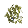
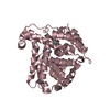
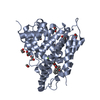
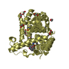
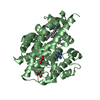
[English] 日本語
Yorodumi
- PDB-1y2d: Catalytic Domain Of Human Phosphodiesterase 4D In Complex With 1-... -

+
Open data


ID or keywords:

Loading...

-
Basic information

Entry
Database: PDB / ID: 1y2d
TitleCatalytic Domain Of Human Phosphodiesterase 4D In Complex With 1-(4-methoxy-phenyl)-3,5-dimethyl-1H-pyrazole-4-carboxylic acid ethyl ester
ComponentscAMP-specific 3',5'-cyclic phosphodiesterase 4D
KeywordsHYDROLASE / Phosphodiesterase / PDE / PDE4D / Pyrazole
Function / homology
Function and homology information


signaling receptor regulator activity / negative regulation of relaxation of cardiac muscle / negative regulation of heart contraction / 3',5'-cyclic-AMP phosphodiesterase / positive regulation of interleukin-5 production / regulation of cardiac muscle cell contraction / establishment of endothelial barrier / negative regulation of cAMP-mediated signaling / beta-2 adrenergic receptor binding / regulation of calcium ion transmembrane transport via high voltage-gated calcium channel ...signaling receptor regulator activity / negative regulation of relaxation of cardiac muscle / negative regulation of heart contraction / 3',5'-cyclic-AMP phosphodiesterase / positive regulation of interleukin-5 production / regulation of cardiac muscle cell contraction / establishment of endothelial barrier / negative regulation of cAMP-mediated signaling / beta-2 adrenergic receptor binding / regulation of calcium ion transmembrane transport via high voltage-gated calcium channel / positive regulation of heart rate / heterocyclic compound binding / adrenergic receptor signaling pathway / voltage-gated calcium channel complex / regulation of cell communication by electrical coupling involved in cardiac conduction / cAMP catabolic process / calcium channel regulator activity / cAMP-mediated signaling / 3',5'-cyclic-nucleotide phosphodiesterase activity / 3',5'-cyclic-AMP phosphodiesterase activity / DARPP-32 events / negative regulation of peptidyl-serine phosphorylation / regulation of release of sequestered calcium ion into cytosol by sarcoplasmic reticulum / cAMP binding / cellular response to cAMP / cellular response to epinephrine stimulus / calcium channel complex / positive regulation of interleukin-2 production / regulation of heart rate / positive regulation of type II interferon production / ATPase binding / T cell receptor signaling pathway / G alpha (s) signalling events / scaffold protein binding / transmembrane transporter binding / apical plasma membrane / centrosome / perinuclear region of cytoplasm / enzyme binding / signal transduction / membrane / metal ion binding / nucleus / plasma membrane / cytosol
Similarity search - Function
Phosphodiesterase 4 upstream conserved regions (UCR) / Phosphodiesterase 4 upstream conserved regions (UCR) / Catalytic domain of cyclic nucleotide phosphodiesterase 4b2b / 3'5'-cyclic nucleotide phosphodiesterase, catalytic domain / 3'5'-cyclic nucleotide phosphodiesterase / 3'5'-cyclic nucleotide phosphodiesterase, catalytic domain / 3'5'-cyclic nucleotide phosphodiesterase, conserved site / 3'5'-cyclic nucleotide phosphodiesterase, catalytic domain superfamily / 3'5'-cyclic nucleotide phosphodiesterase / 3'5'-cyclic nucleotide phosphodiesterase domain signature. ...Phosphodiesterase 4 upstream conserved regions (UCR) / Phosphodiesterase 4 upstream conserved regions (UCR) / Catalytic domain of cyclic nucleotide phosphodiesterase 4b2b / 3'5'-cyclic nucleotide phosphodiesterase, catalytic domain / 3'5'-cyclic nucleotide phosphodiesterase / 3'5'-cyclic nucleotide phosphodiesterase, catalytic domain / 3'5'-cyclic nucleotide phosphodiesterase, conserved site / 3'5'-cyclic nucleotide phosphodiesterase, catalytic domain superfamily / 3'5'-cyclic nucleotide phosphodiesterase / 3'5'-cyclic nucleotide phosphodiesterase domain signature. / 3'5'-cyclic nucleotide phosphodiesterase domain profile. / Orthogonal Bundle / Mainly Alpha
Similarity search - Domain/homology
Chem-4DE / 3',5'-cyclic-AMP phosphodiesterase 4D
Similarity search - Component
Biological speciesHomo sapiens (human)
MethodX-RAY DIFFRACTION / SYNCHROTRON / MOLECULAR REPLACEMENT / Resolution: 1.7 Å
AuthorsCard, G.L. / Blasdel, L. / England, B.P. / Zhang, C. / Suzuki, Y. / Gillette, S. / Fong, D. / Ibrahim, P.N. / Artis, D.R. / Bollag, G. ...Card, G.L. / Blasdel, L. / England, B.P. / Zhang, C. / Suzuki, Y. / Gillette, S. / Fong, D. / Ibrahim, P.N. / Artis, D.R. / Bollag, G. / Milburn, M.V. / Kim, S.-H. / Schlessinger, J. / Zhang, K.Y.J.
CitationJournal: Nat.Biotechnol. / Year: 2005
Title: A family of phosphodiesterase inhibitors discovered by cocrystallography and scaffold-based drug design
Authors: Card, G.L. / Blasdel, L. / England, B.P. / Zhang, C. / Suzuki, Y. / Gillette, S. / Fong, D. / Ibrahim, P.N. / Artis, D.R. / Bollag, G. / Milburn, M.V. / Kim, S.-H. / Schlessinger, J. / Zhang, K.Y.J.
History
DepositionNov 22, 2004Deposition site: RCSB / Processing site: RCSB
Revision 1.0Mar 1, 2005Provider: repository / Type: Initial release
Revision 1.1Apr 30, 2008Group: Version format compliance
Revision 1.2Jul 13, 2011Group: Advisory / Version format compliance
Revision 1.3Feb 14, 2024Group: Advisory / Data collection ...Advisory / Data collection / Database references / Derived calculations / Refinement description
Category: chem_comp_atom / chem_comp_bond ...chem_comp_atom / chem_comp_bond / database_2 / pdbx_database_remark / pdbx_struct_conn_angle / struct_conn / struct_ncs_dom_lim / struct_ref_seq_dif / struct_site
Item: _database_2.pdbx_DOI / _database_2.pdbx_database_accession ..._database_2.pdbx_DOI / _database_2.pdbx_database_accession / _pdbx_database_remark.text / _pdbx_struct_conn_angle.ptnr1_auth_seq_id / _pdbx_struct_conn_angle.ptnr3_auth_seq_id / _pdbx_struct_conn_angle.value / _struct_conn.pdbx_dist_value / _struct_conn.ptnr1_auth_asym_id / _struct_conn.ptnr1_auth_comp_id / _struct_conn.ptnr1_auth_seq_id / _struct_conn.ptnr1_label_asym_id / _struct_conn.ptnr1_label_atom_id / _struct_conn.ptnr1_label_comp_id / _struct_conn.ptnr1_label_seq_id / _struct_conn.ptnr2_auth_asym_id / _struct_conn.ptnr2_auth_comp_id / _struct_conn.ptnr2_auth_seq_id / _struct_conn.ptnr2_label_asym_id / _struct_conn.ptnr2_label_atom_id / _struct_conn.ptnr2_label_comp_id / _struct_ncs_dom_lim.beg_auth_comp_id / _struct_ncs_dom_lim.end_auth_comp_id / _struct_ref_seq_dif.details / _struct_site.pdbx_auth_asym_id / _struct_site.pdbx_auth_comp_id / _struct_site.pdbx_auth_seq_id
Remark 600HETEROGEN HOH 1003-1008 ARE ASSOCIATED WITH CHAIN A. HOH 2003-2008 ARE ASSOCIATED WITH CHAIN B.

-
Structure visualization

Structure viewerMolecule:
MolmilJmol/JSmol

Downloads & links

-
Assembly

Deposited unit
A: cAMP-specific 3',5'-cyclic phosphodiesterase 4D
B: cAMP-specific 3',5'-cyclic phosphodiesterase 4D
hetero molecules


Theoretical massNumber of molelcules
Total (without water)82,69333
Polymers80,1932
Non-polymers2,50031
Water9,728540
1
A: cAMP-specific 3',5'-cyclic phosphodiesterase 4D
hetero molecules


Theoretical massNumber of molelcules
Total (without water)41,20516
Polymers40,0961
Non-polymers1,10915
Water181
TypeNameSymmetry operationNumber
identity operation1_555x,y,z1
2
B: cAMP-specific 3',5'-cyclic phosphodiesterase 4D
hetero molecules


Theoretical massNumber of molelcules
Total (without water)41,48817
Polymers40,0961
Non-polymers1,39116
Water181
TypeNameSymmetry operationNumber
identity operation1_555x,y,z1
Unit cell
Length a, b, c (Å)60.804, 79.146, 164.617
Angle α, β, γ (deg.)90.00, 90.00, 90.00
Int Tables number19
Space group name H-MP212121
Noncrystallographic symmetry (NCS)NCS domain:
IDEns-IDDetails
11A
21B
31A
41B

NCS domain segments:

Ens-ID: 1 / Refine code: 6

Dom-IDComponent-IDBeg auth comp-IDBeg label comp-IDEnd auth comp-IDEnd label comp-IDAuth asym-IDLabel asym-IDAuth seq-IDLabel seq-ID
11GLUGLUVALVALAA87 - 29223 - 228
21GLUGLUVALVALBB87 - 29223 - 228
32VALVALILEILEAA297 - 410233 - 346
42VALVALILEILEBB297 - 410233 - 346
DetailsThe biological assembly is one monomer.

-
Components

-
Protein , 1 types, 2 molecules AB

#1: Protein cAMP-specific 3',5'-cyclic phosphodiesterase 4D / DPDE3 / PDE43


Mass: 40096.355 Da / Num. of mol.: 2 / Fragment: CATALYTIC DOMAIN OF HUMAN PHOSPHODIESTERASE 4D
Source method: isolated from a genetically manipulated source
Source: (gene. exp.) Homo sapiens (human) / Gene: PDE4D / Plasmid: pET15b / Production host: Escherichia coli (E. coli) / Strain (production host): BL21(DE3)Codon Plus(RIL)
References: UniProt: Q08499, 3',5'-cyclic-nucleotide phosphodiesterase

-
Non-polymers , 6 types, 571 molecules

#2: Chemical ChemComp-ZN / ZINC ION


Mass: 65.409 Da / Num. of mol.: 2 / Source method: obtained synthetically / Formula: Zn
#3: Chemical ChemComp-MG / MAGNESIUM ION


Mass: 24.305 Da / Num. of mol.: 2 / Source method: obtained synthetically / Formula: Mg
#4: Chemical ChemComp-4DE / 1-(4-METHOXYPHENYL)-3,5-DIMETHYL-1H-PYRAZOLE-4-CARBOXYLIC ACID ETHYL ESTER


Mass: 274.315 Da / Num. of mol.: 2 / Source method: obtained synthetically / Formula: C15H18N2O3
#5: Chemical...
ChemComp-EDO / 1,2-ETHANEDIOL / ETHYLENE GLYCOL / Ethylene glycol


Mass: 62.068 Da / Num. of mol.: 24 / Source method: obtained synthetically / Formula: C2H6O2
#6: Chemical ChemComp-B3P / 2-[3-(2-HYDROXY-1,1-DIHYDROXYMETHYL-ETHYLAMINO)-PROPYLAMINO]-2-HYDROXYMETHYL-PROPANE-1,3-DIOL / Bis-tris propane


Mass: 282.334 Da / Num. of mol.: 1 / Source method: obtained synthetically / Formula: C11H26N2O6 / Comment: pH buffer*YM
#7: Water ChemComp-HOH / water / Water


Mass: 18.015 Da / Num. of mol.: 540 / Source method: isolated from a natural source / Formula: H2O

-
Experimental details

-
Experiment

ExperimentMethod: X-RAY DIFFRACTION / Number of used crystals: 1

-
Sample preparation

CrystalDensity Matthews: 2.47 Å3/Da / Density % sol: 50.18 %
Crystal growTemperature: 288 K / Method: vapor diffusion, sitting drop / pH: 7
Details: PEG3350, ethylene glycol, isopropanol, glycerol and DTT, pH 7.0, VAPOR DIFFUSION, SITTING DROP, temperature 288K

-
Data collection

DiffractionMean temperature: 93 K
Diffraction sourceSource: SYNCHROTRON / Site: ALS / Beamline: 8.3.1 / Wavelength: 1.1 Å
DetectorType: ADSC QUANTUM 210 / Detector: CCD
RadiationProtocol: SINGLE WAVELENGTH / Monochromatic (M) / Laue (L): M / Scattering type: x-ray
Radiation wavelengthWavelength: 1.1 Å / Relative weight: 1
ReflectionResolution: 1.7→81.65 Å / Num. all: 82929 / Num. obs: 82929 / % possible obs: 99.04 % / Observed criterion σ(F): 0 / Observed criterion σ(I): 0 / Redundancy: 2.9 % / Rmerge(I) obs: 0.066 / Net I/σ(I): 7.6
Reflection shellResolution: 1.7→1.744 Å / Redundancy: 3 % / Rmerge(I) obs: 0.521 / Mean I/σ(I) obs: 1.7 / Num. unique all: 6353 / % possible all: 99.3

-
Processing

Software
NameVersionClassification
REFMAC5.1.25refinement
Blu-Icedata collection
ELVESdata scaling
EPMRphasing
RefinementMethod to determine structure: MOLECULAR REPLACEMENT / Resolution: 1.7→81.65 Å / Cor.coef. Fo:Fc: 0.966 / Cor.coef. Fo:Fc free: 0.955 / SU B: 1.805 / SU ML: 0.058 / TLS residual ADP flag: LIKELY RESIDUAL / Isotropic thermal model: Isotropic / Cross valid method: THROUGHOUT / σ(F): 0 / σ(I): 0 / ESU R: 0.092 / ESU R Free: 0.088 / Stereochemistry target values: MAXIMUM LIKELIHOOD / Details: HYDROGENS HAVE BEEN ADDED IN THE RIDING POSITIONS
RfactorNum. reflection% reflectionSelection details
Rfree0.18908 4382 5 %RANDOM
Rwork0.16525 ---
all0.16646 82929 --
obs0.16646 82929 99.04 %-
Solvent computationIon probe radii: 0.8 Å / Shrinkage radii: 0.8 Å / VDW probe radii: 1.4 Å / Solvent model: BABINET MODEL WITH MASK
Displacement parametersBiso mean: 13.031 Å2
Baniso -1Baniso -2Baniso -3
1-0.51 Å20 Å20 Å2
2--0.56 Å20 Å2
3----1.06 Å2
Refine analyze
FreeObs
Luzzati coordinate error0.088 Å0.092 Å
Refinement stepCycle: LAST / Resolution: 1.7→81.65 Å
ProteinNucleic acidLigandSolventTotal
Num. atoms5276 0 159 540 5975
Refine LS restraints
Refine-IDTypeDev idealDev ideal targetNumber
X-RAY DIFFRACTIONr_bond_refined_d0.010.0215545
X-RAY DIFFRACTIONr_bond_other_d0.0020.024923
X-RAY DIFFRACTIONr_angle_refined_deg1.271.9567480
X-RAY DIFFRACTIONr_angle_other_deg0.865311479
X-RAY DIFFRACTIONr_dihedral_angle_1_deg3.0925650
X-RAY DIFFRACTIONr_dihedral_angle_2_deg
X-RAY DIFFRACTIONr_dihedral_angle_3_deg
X-RAY DIFFRACTIONr_dihedral_angle_4_deg
X-RAY DIFFRACTIONr_chiral_restr0.0810.2853
X-RAY DIFFRACTIONr_gen_planes_refined0.0050.025995
X-RAY DIFFRACTIONr_gen_planes_other0.0050.021033
X-RAY DIFFRACTIONr_nbd_refined0.2150.21237
X-RAY DIFFRACTIONr_nbd_other0.2330.25327
X-RAY DIFFRACTIONr_nbtor_refined
X-RAY DIFFRACTIONr_nbtor_other0.0830.22608
X-RAY DIFFRACTIONr_xyhbond_nbd_refined0.1530.2380
X-RAY DIFFRACTIONr_xyhbond_nbd_other
X-RAY DIFFRACTIONr_metal_ion_refined
X-RAY DIFFRACTIONr_metal_ion_other
X-RAY DIFFRACTIONr_symmetry_vdw_refined0.2220.26
X-RAY DIFFRACTIONr_symmetry_vdw_other0.3270.252
X-RAY DIFFRACTIONr_symmetry_hbond_refined0.1140.219
X-RAY DIFFRACTIONr_symmetry_hbond_other
X-RAY DIFFRACTIONr_mcbond_it0.5721.53268
X-RAY DIFFRACTIONr_mcbond_other
X-RAY DIFFRACTIONr_mcangle_it1.12425336
X-RAY DIFFRACTIONr_scbond_it2.03432277
X-RAY DIFFRACTIONr_scangle_it3.3754.52144
X-RAY DIFFRACTIONr_rigid_bond_restr
X-RAY DIFFRACTIONr_sphericity_free
X-RAY DIFFRACTIONr_sphericity_bonded
Refine LS restraints NCS

Dom-ID: 1 / Auth asym-ID: A / Ens-ID: 1 / Number: 4973 / Refine-ID: X-RAY DIFFRACTION

TypeRms dev position (Å)Weight position
loose positional0.385
loose thermal1.510
LS refinement shellResolution: 1.7→1.744 Å / Total num. of bins used: 20
RfactorNum. reflection% reflection
Rfree0.306 308 -
Rwork0.311 6045 -
obs-6353 99.3 %
Refinement TLS params.

Method: refined / Refine-ID: X-RAY DIFFRACTION

IDL112)L122)L132)L222)L232)L332)S11 (Å °)S12 (Å °)S13 (Å °)S21 (Å °)S22 (Å °)S23 (Å °)S31 (Å °)S32 (Å °)S33 (Å °)T112)T122)T132)T222)T232)T332)Origin x (Å)Origin y (Å)Origin z (Å)
10.67010.1925-0.29790.6421-0.00871.2764-0.06090.0434-0.0123-0.01340.01890.05370.1041-0.07190.0420.065-0.01690.00370.0240.00690.06133.455941.60115.3882
20.43710.0599-0.03140.7143-0.31021.29620.01160.0221-0.0445-0.0096-0.016-0.0668-0.02330.04850.00440.0056-0.00420.01760.0298-0.01220.054715.48943.164754.3542
Refinement TLS group
IDRefine-IDRefine TLS-IDAuth asym-IDLabel asym-IDAuth seq-IDLabel seq-ID
1X-RAY DIFFRACTION1AA86 - 41122 - 347
2X-RAY DIFFRACTION2BB86 - 41122 - 347

+
About Yorodumi

-
News

-
Feb 9, 2022. New format data for meta-information of EMDB entries

New format data for meta-information of EMDB entries

  • Version 3 of the EMDB header file is now the official format.
  • The previous official version 1.9 will be removed from the archive.

Related info.:EMDB header

External links:wwPDB to switch to version 3 of the EMDB data model

-
Aug 12, 2020. Covid-19 info

Covid-19 info

URL: https://pdbj.org/emnavi/covid19.php

New page: Covid-19 featured information page in EM Navigator.

Related info.:Covid-19 info / Mar 5, 2020. Novel coronavirus structure data

+
Mar 5, 2020. Novel coronavirus structure data

Novel coronavirus structure data

Related info.:Yorodumi Speices / Aug 12, 2020. Covid-19 info

External links:COVID-19 featured content - PDBj / Molecule of the Month (242):Coronavirus Proteases

+
Jan 31, 2019. EMDB accession codes are about to change! (news from PDBe EMDB page)

EMDB accession codes are about to change! (news from PDBe EMDB page)

  • The allocation of 4 digits for EMDB accession codes will soon come to an end. Whilst these codes will remain in use, new EMDB accession codes will include an additional digit and will expand incrementally as the available range of codes is exhausted. The current 4-digit format prefixed with “EMD-” (i.e. EMD-XXXX) will advance to a 5-digit format (i.e. EMD-XXXXX), and so on. It is currently estimated that the 4-digit codes will be depleted around Spring 2019, at which point the 5-digit format will come into force.
  • The EM Navigator/Yorodumi systems omit the EMD- prefix.

Related info.:Q: What is EMD? / ID/Accession-code notation in Yorodumi/EM Navigator

External links:EMDB Accession Codes are Changing Soon! / Contact to PDBj

+
Jul 12, 2017. Major update of PDB

Major update of PDB

  • wwPDB released updated PDB data conforming to the new PDBx/mmCIF dictionary.
  • This is a major update changing the version number from 4 to 5, and with Remediation, in which all the entries are updated.
  • In this update, many items about electron microscopy experimental information are reorganized (e.g. em_software).
  • Now, EM Navigator and Yorodumi are based on the updated data.

External links:wwPDB Remediation / Enriched Model Files Conforming to OneDep Data Standards Now Available in the PDB FTP Archive

-
Yorodumi

Thousand views of thousand structures

  • Yorodumi is a browser for structure data from EMDB, PDB, SASBDB, etc.
  • This page is also the successor to EM Navigator detail page, and also detail information page/front-end page for Omokage search.
  • The word "yorodu" (or yorozu) is an old Japanese word meaning "ten thousand". "mi" (miru) is to see.

Related info.:EMDB / PDB / SASBDB / Comparison of 3 databanks / Yorodumi Search / Aug 31, 2016. New EM Navigator & Yorodumi / Yorodumi Papers / Jmol/JSmol / Function and homology information / Changes in new EM Navigator and Yorodumi

Read more