 Movie
Movie Controller

[English] 日本語
 Yorodumi
- PDB-1ptw: The Crystal Structure of AMP-Bound PDE4 Suggests a Mechanism for ... -
+ Open data

- Basic information
| Entry | Database: PDB / ID: 1ptw | ||||||
|---|---|---|---|---|---|---|---|
| Title | The Crystal Structure of AMP-Bound PDE4 Suggests a Mechanism for Phosphodiesterase Catalysis | ||||||
 Components Components | cAMP-specific phosphodiesterase PDE4D2 | ||||||
 Keywords Keywords | HYDROLASE / catalytic mechanism / cAMP hydrolysis crystal structure / binuclear catalysis | ||||||
| Function / homology |  Function and homology information Function and homology informationsignaling receptor regulator activity / negative regulation of relaxation of cardiac muscle / negative regulation of heart contraction / 3',5'-cyclic-AMP phosphodiesterase / positive regulation of interleukin-5 production / negative regulation of adenylate cyclase-activating G protein-coupled receptor signaling pathway / regulation of cardiac muscle cell contraction / establishment of endothelial barrier / regulation of calcium ion transmembrane transport via high voltage-gated calcium channel / heterocyclic compound binding ...signaling receptor regulator activity / negative regulation of relaxation of cardiac muscle / negative regulation of heart contraction / 3',5'-cyclic-AMP phosphodiesterase / positive regulation of interleukin-5 production / negative regulation of adenylate cyclase-activating G protein-coupled receptor signaling pathway / regulation of cardiac muscle cell contraction / establishment of endothelial barrier / regulation of calcium ion transmembrane transport via high voltage-gated calcium channel / heterocyclic compound binding / beta-2 adrenergic receptor binding / voltage-gated calcium channel complex / adrenergic receptor signaling pathway / cAMP catabolic process / 3',5'-cyclic-nucleotide phosphodiesterase activity / 3',5'-cyclic-GMP phosphodiesterase activity / regulation of cell communication by electrical coupling involved in cardiac conduction / 3',5'-cyclic-AMP phosphodiesterase activity / DARPP-32 events / cAMP binding / positive regulation of heart rate / negative regulation of cAMP/PKA signal transduction / regulation of release of sequestered calcium ion into cytosol by sarcoplasmic reticulum / positive regulation of interleukin-2 production / cellular response to epinephrine stimulus / calcium channel complex / regulation of heart rate / Turbulent (oscillatory, disturbed) flow shear stress activates signaling by PIEZO1 and integrins in endothelial cells / cellular response to cAMP / calcium channel regulator activity / positive regulation of type II interferon production / T cell receptor signaling pathway / ATPase binding / nuclear membrane / scaffold protein binding / G alpha (s) signalling events / transmembrane transporter binding / cilium / apical plasma membrane / centrosome / enzyme binding / nucleoplasm / membrane / metal ion binding / plasma membrane / cytosol Similarity search - Function | ||||||
| Biological species |  Homo sapiens (human) Homo sapiens (human) | ||||||
| Method |  X-RAY DIFFRACTION / SYNCHROTRON / MOLECULAR REPLACEMENT / Resolution: 2.3 Å X-RAY DIFFRACTION / SYNCHROTRON / MOLECULAR REPLACEMENT / Resolution: 2.3 Å | ||||||
 Authors Authors | Huai, Q. / Colicelli, J. / Ke, H. | ||||||
 Citation Citation | Journal: Biochemistry / Year: 2003 Title: The crystal structure of AMP-bound PDE4 suggests a mechanism for phosphodiesterase catalysis Authors: Huai, Q. / Colicelli, J. / Ke, H. | ||||||
| History |
| ||||||
| Remark 300 | BIOMOLECULE: 1 THIS ENTRY CONTAINS THE CRYSTALLOGRAPHIC ASYMMETRIC UNIT WHICH CONSISTS OF 4 CHAINS. ...BIOMOLECULE: 1 THIS ENTRY CONTAINS THE CRYSTALLOGRAPHIC ASYMMETRIC UNIT WHICH CONSISTS OF 4 CHAINS. THE BIOLOGICAL UNIT IS UNKNOWN. |
- Structure visualization
| Structure viewer | Molecule: MolmilJmol/JSmol |
|---|
- Downloads & links
-Download
| PDBx/mmCIF format | 1ptw.cif.gz | 274.3 KB | Display | PDBx/mmCIF format |
|---|---|---|---|---|
| PDB format | pdb1ptw.ent.gz | 220.5 KB | Display | PDB format |
| PDBx/mmJSON format | 1ptw.json.gz | Tree view | PDBx/mmJSON format | |
| Others |  Other downloads Other downloads |
-Validation report
| Arichive directory | https://data.pdbj.org/pub/pdb/validation_reports/pt/1ptwftp://data.pdbj.org/pub/pdb/validation_reports/pt/1ptw | HTTPS FTP |
|---|
-Related structure data
| Similar structure data |
|---|


-Links
 PDBj
PDBj


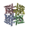
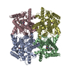
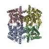
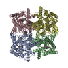
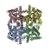
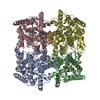
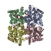
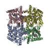
- Assembly
| Deposited unit | 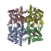
| ||||||||
|---|---|---|---|---|---|---|---|---|---|
| 1 |
| ||||||||
| Unit cell |
|
-Components
| #1: Protein | Mass: 41394.773 Da / Num. of mol.: 4 / Fragment: catalytic domain Source method: isolated from a genetically manipulated source Source: (gene. exp.) Homo sapiens (human) / Gene: PDE4D2 / Plasmid: pET-15b / Species (production host): Escherichia coli / Production host:  References: UniProt: Q08499, 3',5'-cyclic-nucleotide phosphodiesterase #2: Chemical | ChemComp-ZN /   Mass: 65.409 Da / Num. of mol.: 8 / Source method: obtained synthetically / Formula: Zn Mass: 65.409 Da / Num. of mol.: 8 / Source method: obtained synthetically / Formula: Zn#3: Chemical | ChemComp-AMP /   Mass: 347.221 Da / Num. of mol.: 4 / Source method: obtained synthetically / Formula: C10H14N5O7P / Comment: AMP*YM Mass: 347.221 Da / Num. of mol.: 4 / Source method: obtained synthetically / Formula: C10H14N5O7P / Comment: AMP*YM#4: Water | ChemComp-HOH / |  Mass: 18.015 Da / Num. of mol.: 86 / Source method: isolated from a natural source / Formula: H2O Mass: 18.015 Da / Num. of mol.: 86 / Source method: isolated from a natural source / Formula: H2O |
|---|
-Experimental details
-Experiment
| Experiment | Method: X-RAY DIFFRACTION / Number of used crystals: 1 |
|---|
- Sample preparation
| Crystal | Density Matthews: 2.66 Å3/Da / Density % sol: 53.74 % | ||||||||||||||||||||||||||||||||||||||||||||||||||||||||||||||||||||||||||||||||||||
|---|---|---|---|---|---|---|---|---|---|---|---|---|---|---|---|---|---|---|---|---|---|---|---|---|---|---|---|---|---|---|---|---|---|---|---|---|---|---|---|---|---|---|---|---|---|---|---|---|---|---|---|---|---|---|---|---|---|---|---|---|---|---|---|---|---|---|---|---|---|---|---|---|---|---|---|---|---|---|---|---|---|---|---|---|---|
| Crystal grow | Temperature: 277 K / Method: vapor diffusion, hanging drop / pH: 7.5 Details: 50 mM HEPES, 15% PEG 3350, 25% ethylene glycol, 5% methanol, 5% DMSO, pH 7.5, VAPOR DIFFUSION, HANGING DROP, temperature 277K | ||||||||||||||||||||||||||||||||||||||||||||||||||||||||||||||||||||||||||||||||||||
| Crystal grow | *PLUS Temperature: 4 ℃ / Method: vapor diffusion, hanging drop | ||||||||||||||||||||||||||||||||||||||||||||||||||||||||||||||||||||||||||||||||||||
| Components of the solutions | *PLUS
|
-Data collection
| Diffraction | Mean temperature: 100 K |
|---|---|
| Diffraction source | Source: SYNCHROTRON / Site: APS  / Beamline: 14-BM-C / Wavelength: 0.9 Å / Beamline: 14-BM-C / Wavelength: 0.9 Å |
| Detector | Type: ADSC QUANTUM 4 / Detector: CCD / Date: Aug 8, 2002 / Details: mirrors |
| Radiation | Protocol: SINGLE WAVELENGTH / Monochromatic (M) / Laue (L): M / Scattering type: x-ray |
| Radiation wavelength | Wavelength: 0.9 Å / Relative weight: 1 |
| Reflection | Resolution: 2.3→99 Å / Num. obs: 67136 / Observed criterion σ(F): 0 / Observed criterion σ(I): 0 / Redundancy: 6 % / Rsym value: 0.075 / Net I/σ(I): 21.9 |
| Reflection shell | Resolution: 2.3→2.38 Å / Rmerge(I) obs: 0.539 / Mean I/σ(I) obs: 3.9 / Num. unique all: 5658 / % possible all: 72.6 |
| Reflection | *PLUS Highest resolution: 2.4 Å / Num. obs: 64052 / % possible obs: 91.8 % / Num. measured all: 404282 / Rmerge(I) obs: 0.075 |
| Reflection shell | *PLUS Highest resolution: 2.4 Å / Lowest resolution: 2.5 Å / % possible obs: 87.9 % / Rmerge(I) obs: 0.45 / Mean I/σ(I) obs: 5.3 |
- Processing
| Software |
| ||||||||||||||||||||||||||||
|---|---|---|---|---|---|---|---|---|---|---|---|---|---|---|---|---|---|---|---|---|---|---|---|---|---|---|---|---|---|
| Refinement | Method to determine structure: MOLECULAR REPLACEMENT / Resolution: 2.3→99 Å / Isotropic thermal model: isotropic / Cross valid method: THROUGHOUT / σ(F): 0 / σ(I): 0 / Stereochemistry target values: Engh & Huber
| ||||||||||||||||||||||||||||
| Displacement parameters |
| ||||||||||||||||||||||||||||
| Refinement step | Cycle: LAST / Resolution: 2.3→99 Å
| ||||||||||||||||||||||||||||
| Refine LS restraints |
| ||||||||||||||||||||||||||||
| Xplor file |
| ||||||||||||||||||||||||||||
| Refinement | *PLUS Highest resolution: 2.4 Å | ||||||||||||||||||||||||||||
| Solvent computation | *PLUS | ||||||||||||||||||||||||||||
| Displacement parameters | *PLUS |
