 Movie
Movie Controller

[English] 日本語
 Yorodumi
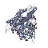
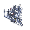
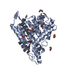
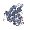
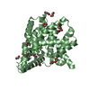
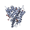
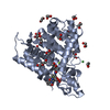
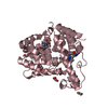
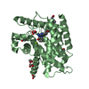
- PDB-3sl4: Crystal structure of the catalytic domain of PDE4D2 with compound 10D -
+ Open data

- Basic information
| Entry | Database: PDB / ID: 3sl4 | ||||||
|---|---|---|---|---|---|---|---|
| Title | Crystal structure of the catalytic domain of PDE4D2 with compound 10D | ||||||
 Components Components | cAMP-specific 3',5'-cyclic phosphodiesterase 4D | ||||||
 Keywords Keywords | Hydrolase/Inhibitor / catalytic mechanism / cAMP hydrolysis / Hydrolase-Inhibitor complex | ||||||
| Function / homology |  Function and homology information Function and homology informationsignaling receptor regulator activity / negative regulation of relaxation of cardiac muscle / negative regulation of heart contraction / 3',5'-cyclic-AMP phosphodiesterase / positive regulation of interleukin-5 production / negative regulation of adenylate cyclase-activating G protein-coupled receptor signaling pathway / regulation of cardiac muscle cell contraction / establishment of endothelial barrier / regulation of calcium ion transmembrane transport via high voltage-gated calcium channel / heterocyclic compound binding ...signaling receptor regulator activity / negative regulation of relaxation of cardiac muscle / negative regulation of heart contraction / 3',5'-cyclic-AMP phosphodiesterase / positive regulation of interleukin-5 production / negative regulation of adenylate cyclase-activating G protein-coupled receptor signaling pathway / regulation of cardiac muscle cell contraction / establishment of endothelial barrier / regulation of calcium ion transmembrane transport via high voltage-gated calcium channel / heterocyclic compound binding / beta-2 adrenergic receptor binding / voltage-gated calcium channel complex / adrenergic receptor signaling pathway / cAMP catabolic process / 3',5'-cyclic-nucleotide phosphodiesterase activity / 3',5'-cyclic-GMP phosphodiesterase activity / regulation of cell communication by electrical coupling involved in cardiac conduction / 3',5'-cyclic-AMP phosphodiesterase activity / DARPP-32 events / positive regulation of interleukin-2 production / cAMP binding / positive regulation of heart rate / negative regulation of cAMP/PKA signal transduction / regulation of release of sequestered calcium ion into cytosol by sarcoplasmic reticulum / cellular response to epinephrine stimulus / calcium channel complex / regulation of heart rate / Turbulent (oscillatory, disturbed) flow shear stress activates signaling by PIEZO1 and integrins in endothelial cells / cellular response to cAMP / calcium channel regulator activity / positive regulation of type II interferon production / T cell receptor signaling pathway / ATPase binding / scaffold protein binding / nuclear membrane / G alpha (s) signalling events / transmembrane transporter binding / apical plasma membrane / cilium / centrosome / enzyme binding / nucleoplasm / membrane / metal ion binding / plasma membrane / cytosol Similarity search - Function | ||||||
| Biological species |  Homo sapiens (human) Homo sapiens (human) | ||||||
| Method |  X-RAY DIFFRACTION / SYNCHROTRON / molrep / Resolution: 1.9 Å X-RAY DIFFRACTION / SYNCHROTRON / molrep / Resolution: 1.9 Å | ||||||
 Authors Authors | Feil, S.F. | ||||||
 Citation Citation | Journal: Bioorg.Med.Chem.Lett. / Year: 2011 Title: Thiophene inhibitors of PDE4: Crystal structures show a second binding mode at the catalytic domain of PDE4D2. Authors: Nankervis, J.L. / Feil, S.C. / Hancock, N.C. / Zheng, Z. / Ng, H.L. / Morton, C.J. / Holien, J.K. / Ho, P.W. / Frazzetto, M.M. / Jennings, I.G. / Manallack, D.T. / Martin, T.J. / Thompson, P.E. / Parker, M.W. | ||||||
| History |
|
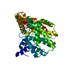
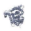
- Structure visualization
| Structure viewer | Molecule: MolmilJmol/JSmol |
|---|
- Downloads & links
-Download
| PDBx/mmCIF format | 3sl4.cif.gz | 297.8 KB | Display | PDBx/mmCIF format |
|---|---|---|---|---|
| PDB format | pdb3sl4.ent.gz | 238.5 KB | Display | PDB format |
| PDBx/mmJSON format | 3sl4.json.gz | Tree view | PDBx/mmJSON format | |
| Others |  Other downloads Other downloads |
-Validation report
| Arichive directory | https://data.pdbj.org/pub/pdb/validation_reports/sl/3sl4ftp://data.pdbj.org/pub/pdb/validation_reports/sl/3sl4 | HTTPS FTP |
|---|
-Related structure data


-Links
 PDBj
PDBj


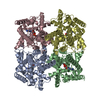
- Assembly
| Deposited unit | 
| ||||||||
|---|---|---|---|---|---|---|---|---|---|
| 1 | 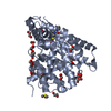
| ||||||||
| 2 | 
| ||||||||
| 3 | 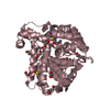
| ||||||||
| 4 | 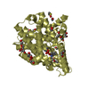
| ||||||||
| Unit cell |
| ||||||||
| Details | AS PER THE AUTHORS THE BIOLOGICAL ASSEMBLY IS UNKNOWN |
-Components
-Protein , 1 types, 4 molecules ABCD
| #1: Protein | Mass: 41523.887 Da / Num. of mol.: 4 / Fragment: Catalytic domain, UNP residues 381-741 Source method: isolated from a genetically manipulated source Source: (gene. exp.) Homo sapiens (human) / Gene: PDE4D, DPDE3 / Plasmid: pET15b / Production host:  References: UniProt: Q08499, 3',5'-cyclic-nucleotide phosphodiesterase |
|---|
-Non-polymers , 8 types, 549 molecules 


| #2: Chemical | ChemComp-ZN /  Mass: 65.409 Da / Num. of mol.: 8 / Source method: obtained synthetically / Formula: Zn Mass: 65.409 Da / Num. of mol.: 8 / Source method: obtained synthetically / Formula: Zn#3: Chemical | ChemComp-EDO /  Mass: 62.068 Da / Num. of mol.: 51 / Source method: obtained synthetically / Formula: C2H6O2 Mass: 62.068 Da / Num. of mol.: 51 / Source method: obtained synthetically / Formula: C2H6O2#4: Chemical | ChemComp-DMS /  Mass: 78.133 Da / Num. of mol.: 8 / Source method: obtained synthetically / Formula: C2H6OS / Comment: DMSO, precipitant*YM Mass: 78.133 Da / Num. of mol.: 8 / Source method: obtained synthetically / Formula: C2H6OS / Comment: DMSO, precipitant*YM#5: Chemical | ChemComp-PEG /  Mass: 106.120 Da / Num. of mol.: 5 / Source method: obtained synthetically / Formula: C4H10O3 Mass: 106.120 Da / Num. of mol.: 5 / Source method: obtained synthetically / Formula: C4H10O3#6: Chemical | ChemComp-PO4 /  Mass: 94.971 Da / Num. of mol.: 4 / Source method: obtained synthetically / Formula: PO4 Mass: 94.971 Da / Num. of mol.: 4 / Source method: obtained synthetically / Formula: PO4#7: Chemical | ChemComp-JN4 /  Mass: 411.474 Da / Num. of mol.: 4 / Source method: obtained synthetically / Formula: C21H21N3O4S Mass: 411.474 Da / Num. of mol.: 4 / Source method: obtained synthetically / Formula: C21H21N3O4S#8: Chemical |  Mass: 238.305 Da / Num. of mol.: 2 / Source method: obtained synthetically / Formula: C8H18N2O4S / Comment: pH buffer*YM Mass: 238.305 Da / Num. of mol.: 2 / Source method: obtained synthetically / Formula: C8H18N2O4S / Comment: pH buffer*YM#9: Water | ChemComp-HOH / | Mass: 18.015 Da / Num. of mol.: 467 / Source method: isolated from a natural source / Formula: H2O |
|---|
-Experimental details
-Experiment
| Experiment | Method: X-RAY DIFFRACTION / Number of used crystals: 1 |
|---|
- Sample preparation
| Crystal | Density Matthews: 2.93 Å3/Da / Density % sol: 57.99 % |
|---|---|
| Crystal grow | Temperature: 294 K / Method: vapor diffusion, hanging drop / pH: 7.5 Details: 20% PEG 3350, 25% (v/v) ethylene glycol, 10% (v/v) isopropanol, 100 mM HEPES, pH 7.5, vapor diffusion, hanging drop, temperature 294K |
-Data collection
| Diffraction | Mean temperature: 100 K | |||||||||||||||||||||||||||||||||||||||||||||||||||||||||||||||||||||||||||||
|---|---|---|---|---|---|---|---|---|---|---|---|---|---|---|---|---|---|---|---|---|---|---|---|---|---|---|---|---|---|---|---|---|---|---|---|---|---|---|---|---|---|---|---|---|---|---|---|---|---|---|---|---|---|---|---|---|---|---|---|---|---|---|---|---|---|---|---|---|---|---|---|---|---|---|---|---|---|---|
| Diffraction source | Source: SYNCHROTRON / Site: Australian Synchrotron  / Beamline: MX1 / Wavelength: 1 Å / Beamline: MX1 / Wavelength: 1 Å | |||||||||||||||||||||||||||||||||||||||||||||||||||||||||||||||||||||||||||||
| Detector | Type: ADSC QUANTUM 210r / Detector: CCD / Date: Feb 17, 2010 | |||||||||||||||||||||||||||||||||||||||||||||||||||||||||||||||||||||||||||||
| Radiation | Monochromator: K-B pair of biomorph mirrors / Protocol: SINGLE WAVELENGTH / Monochromatic (M) / Laue (L): M / Scattering type: x-ray | |||||||||||||||||||||||||||||||||||||||||||||||||||||||||||||||||||||||||||||
| Radiation wavelength | Wavelength: 1 Å / Relative weight: 1 | |||||||||||||||||||||||||||||||||||||||||||||||||||||||||||||||||||||||||||||
| Reflection | Resolution: 1.9→50 Å / Num. obs: 139220 / % possible obs: 99.1 % / Observed criterion σ(F): 2 / Observed criterion σ(I): 2 / Redundancy: 5 % / Rmerge(I) obs: 0.065 / Χ2: 1.063 / Net I/σ(I): 10.5 | |||||||||||||||||||||||||||||||||||||||||||||||||||||||||||||||||||||||||||||
| Reflection shell |
|
- Processing
| Software |
| |||||||||||||||||||||||||||||||||||||||||||||||||||||||||||||||||||||||||||||||||||||||||||||||||||||||||||||||||||||||||||||||||||||||||||||||||||||||||||||||||||||||||||||||||||||||||||||||||||||||||||||||||||||||||
|---|---|---|---|---|---|---|---|---|---|---|---|---|---|---|---|---|---|---|---|---|---|---|---|---|---|---|---|---|---|---|---|---|---|---|---|---|---|---|---|---|---|---|---|---|---|---|---|---|---|---|---|---|---|---|---|---|---|---|---|---|---|---|---|---|---|---|---|---|---|---|---|---|---|---|---|---|---|---|---|---|---|---|---|---|---|---|---|---|---|---|---|---|---|---|---|---|---|---|---|---|---|---|---|---|---|---|---|---|---|---|---|---|---|---|---|---|---|---|---|---|---|---|---|---|---|---|---|---|---|---|---|---|---|---|---|---|---|---|---|---|---|---|---|---|---|---|---|---|---|---|---|---|---|---|---|---|---|---|---|---|---|---|---|---|---|---|---|---|---|---|---|---|---|---|---|---|---|---|---|---|---|---|---|---|---|---|---|---|---|---|---|---|---|---|---|---|---|---|---|---|---|---|---|---|---|---|---|---|---|---|---|---|---|---|---|---|---|---|
| Refinement | Method to determine structure: molrep / Resolution: 1.9→32.621 Å / Occupancy max: 1 / Occupancy min: 0.08 / FOM work R set: 0.8499 / SU ML: 0.25 / σ(F): 0.05 / Phase error: 22.36 / Stereochemistry target values: ML
| |||||||||||||||||||||||||||||||||||||||||||||||||||||||||||||||||||||||||||||||||||||||||||||||||||||||||||||||||||||||||||||||||||||||||||||||||||||||||||||||||||||||||||||||||||||||||||||||||||||||||||||||||||||||||
| Solvent computation | Shrinkage radii: 0.9 Å / VDW probe radii: 1.11 Å / Solvent model: FLAT BULK SOLVENT MODEL / Bsol: 60.572 Å2 / ksol: 0.4 e/Å3 | |||||||||||||||||||||||||||||||||||||||||||||||||||||||||||||||||||||||||||||||||||||||||||||||||||||||||||||||||||||||||||||||||||||||||||||||||||||||||||||||||||||||||||||||||||||||||||||||||||||||||||||||||||||||||
| Displacement parameters | Biso max: 88.64 Å2 / Biso mean: 32.3233 Å2 / Biso min: 10.87 Å2
| |||||||||||||||||||||||||||||||||||||||||||||||||||||||||||||||||||||||||||||||||||||||||||||||||||||||||||||||||||||||||||||||||||||||||||||||||||||||||||||||||||||||||||||||||||||||||||||||||||||||||||||||||||||||||
| Refinement step | Cycle: LAST / Resolution: 1.9→32.621 Å
| |||||||||||||||||||||||||||||||||||||||||||||||||||||||||||||||||||||||||||||||||||||||||||||||||||||||||||||||||||||||||||||||||||||||||||||||||||||||||||||||||||||||||||||||||||||||||||||||||||||||||||||||||||||||||
| Refine LS restraints |
| |||||||||||||||||||||||||||||||||||||||||||||||||||||||||||||||||||||||||||||||||||||||||||||||||||||||||||||||||||||||||||||||||||||||||||||||||||||||||||||||||||||||||||||||||||||||||||||||||||||||||||||||||||||||||
| LS refinement shell | Refine-ID: X-RAY DIFFRACTION / Total num. of bins used: 30
|
