 Movie
Movie Controller

[English] 日本語
 Yorodumi
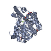
- PDB-6rb6: Crystal structure of T. brucei PDE-B1 catalytic domain with inhib... -
+ Open data

- Basic information
| Entry | Database: PDB / ID: 6rb6 | ||||||
|---|---|---|---|---|---|---|---|
| Title | Crystal structure of T. brucei PDE-B1 catalytic domain with inhibitor NPD-053 | ||||||
 Components Components | Phosphodiesterase | ||||||
 Keywords Keywords | HYDROLASE / Parasitic phosphodiesterase / African trypanosomiasis / sleeping sickness | ||||||
| Function / homology |  Function and homology information Function and homology informationHydrolases; Acting on ester bonds; Phosphoric-diester hydrolases / 3',5'-cyclic-nucleotide phosphodiesterase activity / 3',5'-cyclic-AMP phosphodiesterase activity / axoneme / cell morphogenesis / signal transduction / metal ion binding / cytoplasm Similarity search - Function | ||||||
| Biological species |  | ||||||
| Method |  X-RAY DIFFRACTION / SYNCHROTRON / MOLECULAR REPLACEMENT / Resolution: 1.9 Å X-RAY DIFFRACTION / SYNCHROTRON / MOLECULAR REPLACEMENT / Resolution: 1.9 Å | ||||||
 Authors Authors | Singh, A.K. / Brown, D.G. | ||||||
| Funding support | 1items
| ||||||
 Citation Citation | Journal: Bioorg.Med.Chem. / Year: 2019 Title: Alkynamide phthalazinones as a new class of TbrPDEB1 inhibitors (Part 2). Authors: de Heuvel, E. / Singh, A.K. / Boronat, P. / Kooistra, A.J. / van der Meer, T. / Sadek, P. / Blaazer, A.R. / Shaner, N.C. / Bindels, D.S. / Caljon, G. / Maes, L. / Sterk, G.J. / Siderius, M. ...Authors: de Heuvel, E. / Singh, A.K. / Boronat, P. / Kooistra, A.J. / van der Meer, T. / Sadek, P. / Blaazer, A.R. / Shaner, N.C. / Bindels, D.S. / Caljon, G. / Maes, L. / Sterk, G.J. / Siderius, M. / Oberholzer, M. / de Esch, I.J.P. / Brown, D.G. / Leurs, R. | ||||||
| History |
|
- Structure visualization
| Structure viewer | Molecule: MolmilJmol/JSmol |
|---|
- Downloads & links
-Download
| PDBx/mmCIF format | 6rb6.cif.gz | 162.1 KB | Display | PDBx/mmCIF format |
|---|---|---|---|---|
| PDB format | pdb6rb6.ent.gz | 125.5 KB | Display | PDB format |
| PDBx/mmJSON format | 6rb6.json.gz | Tree view | PDBx/mmJSON format | |
| Others |  Other downloads Other downloads |
-Validation report
| Arichive directory | https://data.pdbj.org/pub/pdb/validation_reports/rb/6rb6ftp://data.pdbj.org/pub/pdb/validation_reports/rb/6rb6 | HTTPS FTP |
|---|
-Related structure data
| Related structure data |  6gxqC  6hwoC  6rcwC  6rfnC  6rfwC  6rgkC 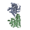 4i15S C: citing same article ( S: Starting model for refinement |
|---|---|
| Similar structure data |

-Links
 PDBj
PDBj

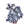
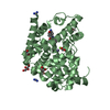
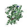
- Assembly
| Deposited unit | 
| ||||||||
|---|---|---|---|---|---|---|---|---|---|
| 1 | 
| ||||||||
| 2 | 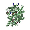
| ||||||||
| Unit cell |
|
-Components
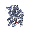
-Protein , 1 types, 2 molecules AB
| #1: Protein | Mass: 40623.340 Da / Num. of mol.: 2 Source method: isolated from a genetically manipulated source Details: residues 565-918 / Source: (gene. exp.)  References: UniProt: Q8WQX9, Hydrolases; Acting on ester bonds; Phosphoric-diester hydrolases |
|---|
-Non-polymers , 7 types, 447 molecules 

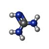


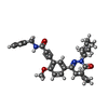

| #2: Chemical |  Mass: 24.305 Da / Num. of mol.: 2 / Source method: obtained synthetically / Formula: Mg Mass: 24.305 Da / Num. of mol.: 2 / Source method: obtained synthetically / Formula: Mg#3: Chemical |  Mass: 65.409 Da / Num. of mol.: 2 / Source method: obtained synthetically / Formula: Zn Mass: 65.409 Da / Num. of mol.: 2 / Source method: obtained synthetically / Formula: Zn#4: Chemical | ChemComp-GAI /  Mass: 59.070 Da / Num. of mol.: 12 / Source method: obtained synthetically / Formula: CH5N3 Mass: 59.070 Da / Num. of mol.: 12 / Source method: obtained synthetically / Formula: CH5N3#5: Chemical | ChemComp-EDO /  Mass: 62.068 Da / Num. of mol.: 9 / Source method: obtained synthetically / Formula: C2H6O2 Mass: 62.068 Da / Num. of mol.: 9 / Source method: obtained synthetically / Formula: C2H6O2#6: Chemical |  Mass: 46.025 Da / Num. of mol.: 2 / Source method: obtained synthetically / Formula: CH2O2 Mass: 46.025 Da / Num. of mol.: 2 / Source method: obtained synthetically / Formula: CH2O2#7: Chemical |  Mass: 499.601 Da / Num. of mol.: 2 / Source method: obtained synthetically / Formula: C30H33N3O4 / Feature type: SUBJECT OF INVESTIGATION Mass: 499.601 Da / Num. of mol.: 2 / Source method: obtained synthetically / Formula: C30H33N3O4 / Feature type: SUBJECT OF INVESTIGATION#8: Water | ChemComp-HOH / | Mass: 18.015 Da / Num. of mol.: 418 / Source method: isolated from a natural source / Formula: H2O |
|---|
-Experimental details
-Experiment
| Experiment | Method: X-RAY DIFFRACTION / Number of used crystals: 1 |
|---|
- Sample preparation
| Crystal | Density Matthews: 3.15 Å3/Da / Density % sol: 61 % |
|---|---|
| Crystal grow | Temperature: 277 K / Method: vapor diffusion, hanging drop / pH: 6.5 Details: 20% PEG 3350, 0.4 M sodium formate, 0.3 M guanidine, 0.1 M MES pH 6.5 |
-Data collection
| Diffraction | Mean temperature: 100 K / Serial crystal experiment: N |
|---|---|
| Diffraction source | Source: SYNCHROTRON / Site: Diamond  / Beamline: I04-1 / Wavelength: 0.92819 Å / Beamline: I04-1 / Wavelength: 0.92819 Å |
| Detector | Type: DECTRIS PILATUS 6M-F / Detector: PIXEL / Date: Aug 3, 2015 |
| Radiation | Protocol: SINGLE WAVELENGTH / Monochromatic (M) / Laue (L): M / Scattering type: x-ray |
| Radiation wavelength | Wavelength: 0.92819 Å / Relative weight: 1 |
| Reflection | Resolution: 1.9→69.57 Å / Num. obs: 78709 / % possible obs: 99.7 % / Redundancy: 3.1 % / CC1/2: 0.995 / Rmerge(I) obs: 0.091 / Rpim(I) all: 0.061 / Rrim(I) all: 0.11 / Net I/σ(I): 7.2 |
| Reflection shell | Resolution: 1.9→1.94 Å / Redundancy: 3.3 % / Rmerge(I) obs: 0.626 / Mean I/σ(I) obs: 2 / Num. unique obs: 4518 / CC1/2: 0.475 / Rpim(I) all: 0.405 / Rrim(I) all: 0.748 / % possible all: 99.9 |
- Processing
| Software |
| ||||||||||||||||||||||||||||||||||||||||||||||||||||||||||||||||||||||||||||||||||||||||||||||||||||||||||||||||||||||||||||||||||||||||||||||||||||||||||||||||||||||||||||||||||||||
|---|---|---|---|---|---|---|---|---|---|---|---|---|---|---|---|---|---|---|---|---|---|---|---|---|---|---|---|---|---|---|---|---|---|---|---|---|---|---|---|---|---|---|---|---|---|---|---|---|---|---|---|---|---|---|---|---|---|---|---|---|---|---|---|---|---|---|---|---|---|---|---|---|---|---|---|---|---|---|---|---|---|---|---|---|---|---|---|---|---|---|---|---|---|---|---|---|---|---|---|---|---|---|---|---|---|---|---|---|---|---|---|---|---|---|---|---|---|---|---|---|---|---|---|---|---|---|---|---|---|---|---|---|---|---|---|---|---|---|---|---|---|---|---|---|---|---|---|---|---|---|---|---|---|---|---|---|---|---|---|---|---|---|---|---|---|---|---|---|---|---|---|---|---|---|---|---|---|---|---|---|---|---|---|
| Refinement | Method to determine structure: MOLECULAR REPLACEMENT Starting model: 4I15 Resolution: 1.9→69.57 Å / Cor.coef. Fo:Fc: 0.967 / Cor.coef. Fo:Fc free: 0.949 / SU B: 3.174 / SU ML: 0.087 / Cross valid method: THROUGHOUT / ESU R: 0.108 / ESU R Free: 0.107 / Details: HYDROGENS HAVE BEEN ADDED IN THE RIDING POSITIONS
| ||||||||||||||||||||||||||||||||||||||||||||||||||||||||||||||||||||||||||||||||||||||||||||||||||||||||||||||||||||||||||||||||||||||||||||||||||||||||||||||||||||||||||||||||||||||
| Solvent computation | Ion probe radii: 0.8 Å / Shrinkage radii: 0.8 Å / VDW probe radii: 1.2 Å | ||||||||||||||||||||||||||||||||||||||||||||||||||||||||||||||||||||||||||||||||||||||||||||||||||||||||||||||||||||||||||||||||||||||||||||||||||||||||||||||||||||||||||||||||||||||
| Displacement parameters | Biso mean: 30.256 Å2
| ||||||||||||||||||||||||||||||||||||||||||||||||||||||||||||||||||||||||||||||||||||||||||||||||||||||||||||||||||||||||||||||||||||||||||||||||||||||||||||||||||||||||||||||||||||||
| Refinement step | Cycle: 1 / Resolution: 1.9→69.57 Å
| ||||||||||||||||||||||||||||||||||||||||||||||||||||||||||||||||||||||||||||||||||||||||||||||||||||||||||||||||||||||||||||||||||||||||||||||||||||||||||||||||||||||||||||||||||||||
| Refine LS restraints |
|