 Movie
Movie Controller

[English] 日本語
 Yorodumi
- PDB-3six: Crystal structure of NodZ alpha-1,6-fucosyltransferase soaked wit... -
+ Open data

- Basic information
| Entry | Database: PDB / ID: 3six | ||||||
|---|---|---|---|---|---|---|---|
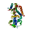
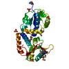
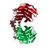
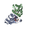
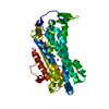
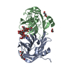
| Title | Crystal structure of NodZ alpha-1,6-fucosyltransferase soaked with GDP-fucose | ||||||
 Components Components | Nodulation fucosyltransferase NodZ | ||||||
 Keywords Keywords | TRANSFERASE / family GT23 glycosyltransferase / GT-B fold / alfa1 / 6-fucosyltransferase / nodulation protein / chitooligosaccharide fucosylation / Nod Factor biosynthesis / nitrogen fixation / legume-rhizobium symbiosis | ||||||
| Function / homology |  Function and homology information Function and homology information | ||||||
| Biological species |  Bradyrhizobium sp. (bacteria) Bradyrhizobium sp. (bacteria) | ||||||
| Method |  X-RAY DIFFRACTION / SYNCHROTRON / MOLECULAR REPLACEMENT / Resolution: 2.35 Å X-RAY DIFFRACTION / SYNCHROTRON / MOLECULAR REPLACEMENT / Resolution: 2.35 Å | ||||||
 Authors Authors | Brzezinski, K. / Dauter, Z. / Jaskolski, M. | ||||||
 Citation Citation | Journal: Acta Crystallogr.,Sect.D / Year: 2012 Title: Structures of NodZ alpha-1,6-fucosyltransferase in complex with GDP and GDP-fucose Authors: Brzezinski, K. / Dauter, Z. / Jaskolski, M. #1: Journal: Acta Biochim.Pol. / Year: 2007Title: High-resolution structure of NodZ fucosyltransferase involved in the biosynthesis of the nodulation factor Authors: Brzezinski, K. / Stepkowski, T. / Panjikar, S. / Bujacz, G. / Jaskolski, M. #2: Journal: J.Biol.Chem. / Year: 2007Title: Structure and Mechanism of Helicobacter pylori Fucosyltransferase: A basis for lipopolysaccharide variation and inhibitor design Authors: Sun, H.Y. / Lin, S.W. / Ko, T.P. / Liu, C.L. / Wang, H.J. / Lin, C.H. #3: Journal: Glycobiology / Year: 2007Title: Crystal structure of human alpha 1,6-fucosyltransferase, FUT8 Authors: Ihara, H. / Ikeda, Y. / Toma, S. / Wang, X. / Suzuki, T. / Gu, J. / Miyoshi, E. / Tsukihara, T. / Honke, K. / Matsumoto, A. / Nakagawa, A. / Taniguchi, N. #4: Journal: Acta Crystallogr.,Sect.D / Year: 2004Title: Cloning, purification, crystallization and preliminary crystallographic studies of bradyrhizobium fucosyltransferase NodZ Authors: Brzezinski, K. / Rogozinski, B. / Stepkowski, T. / Bujacz, G. / Jaskolski, M. #5: Journal: Proc.Natl.Acad.Sci.USA / Year: 1997Title: Bacterial nodulation protein NodZ is a chitin oligosaccharide fucosyltransferase which can also recognize related substrates of animal origin Authors: Quinto, C. / Wijfjes, A.H. / Bloemberg, G.V. / Blok-Tip, L. / Lopez-Lara, I.M. / Lugtenberg, B.J. / Thomas-Oates, J.E. / Spaink, H.P. #6: Journal: J. Bacteriol. / Year: 1997Title: Rhizobium sp. strain NGR234 NodZ protein is a fucosyltransferase Authors: Quesada-Vincens, D. / Fellay, R. / Nasim, T. / Viprey, V. / Burger, U. / Prome, J.C. / Broughton, W.J. / Jabbouri, S. | ||||||
| History |
|
- Structure visualization
| Structure viewer | Molecule: MolmilJmol/JSmol |
|---|
- Downloads & links
-Download
| PDBx/mmCIF format | 3six.cif.gz | 138.7 KB | Display | PDBx/mmCIF format |
|---|---|---|---|---|
| PDB format | pdb3six.ent.gz | 108.2 KB | Display | PDB format |
| PDBx/mmJSON format | 3six.json.gz | Tree view | PDBx/mmJSON format | |
| Others |  Other downloads Other downloads |
-Validation report
| Arichive directory | https://data.pdbj.org/pub/pdb/validation_reports/si/3sixftp://data.pdbj.org/pub/pdb/validation_reports/si/3six | HTTPS FTP |
|---|
-Related structure data
| Related structure data | 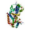 3siwC 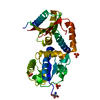 2hhcS S: Starting model for refinement C: citing same article ( |
|---|---|
| Similar structure data |


-Links
 PDBj
PDBj- Assembly
| Deposited unit | 
| ||||||||
|---|---|---|---|---|---|---|---|---|---|
| 1 |
| ||||||||
| 2 | 
| ||||||||
| Unit cell |
|
-Components
| #1: Protein | Mass: 37828.988 Da / Num. of mol.: 1 Source method: isolated from a genetically manipulated source Source: (gene. exp.) Bradyrhizobium sp. (bacteria) / Strain: WM9 / Gene: nodZ / Plasmid: pET3a / Production host: References: UniProt: Q9AQ17, Transferases; Glycosyltransferases; Hexosyltransferases |
|---|---|
| #2: Chemical | ChemComp-GDP /   Type: RNA linking / Mass: 443.201 Da / Num. of mol.: 1 / Source method: obtained synthetically / Formula: C10H15N5O11P2 / Comment: GDP, energy-carrying molecule*YM Type: RNA linking / Mass: 443.201 Da / Num. of mol.: 1 / Source method: obtained synthetically / Formula: C10H15N5O11P2 / Comment: GDP, energy-carrying molecule*YM |
| #3: Chemical | ChemComp-PO4 /   Mass: 94.971 Da / Num. of mol.: 1 / Source method: obtained synthetically / Formula: PO4 Mass: 94.971 Da / Num. of mol.: 1 / Source method: obtained synthetically / Formula: PO4 |
| #4: Chemical | ChemComp-CL /   Mass: 35.453 Da / Num. of mol.: 1 / Source method: obtained synthetically / Formula: Cl Mass: 35.453 Da / Num. of mol.: 1 / Source method: obtained synthetically / Formula: Cl |
| #5: Water | ChemComp-HOH /  Mass: 18.015 Da / Num. of mol.: 82 / Source method: isolated from a natural source / Formula: H2O Mass: 18.015 Da / Num. of mol.: 82 / Source method: isolated from a natural source / Formula: H2O |
-Experimental details
-Experiment
| Experiment | Method: X-RAY DIFFRACTION / Number of used crystals: 1 |
|---|
- Sample preparation
| Crystal | Density Matthews: 2.88 Å3/Da / Density % sol: 57.34 % |
|---|---|
| Crystal grow | Temperature: 293 K / Method: vapor diffusion, hanging drop / pH: 6.5 Details: 350 mM potassium sodium tartrate 100 mM MES pH 6.5 50 mM MgCl2 , VAPOR DIFFUSION, HANGING DROP, temperature 293K |
-Data collection
| Diffraction | Mean temperature: 100 K | |||||||||||||||||||||||||||||||||||||||||||||||||||||||||||||||||||||||||||||
|---|---|---|---|---|---|---|---|---|---|---|---|---|---|---|---|---|---|---|---|---|---|---|---|---|---|---|---|---|---|---|---|---|---|---|---|---|---|---|---|---|---|---|---|---|---|---|---|---|---|---|---|---|---|---|---|---|---|---|---|---|---|---|---|---|---|---|---|---|---|---|---|---|---|---|---|---|---|---|
| Diffraction source | Source: SYNCHROTRON / Site: APS  / Beamline: 22-BM / Wavelength: 0.97242 Å / Beamline: 22-BM / Wavelength: 0.97242 Å | |||||||||||||||||||||||||||||||||||||||||||||||||||||||||||||||||||||||||||||
| Detector | Type: MARMOSAIC 225 mm CCD / Detector: CCD / Date: Apr 24, 2011 | |||||||||||||||||||||||||||||||||||||||||||||||||||||||||||||||||||||||||||||
| Radiation | Monochromator: double crystal Si(220) / Protocol: SINGLE WAVELENGTH / Monochromatic (M) / Laue (L): M / Scattering type: x-ray | |||||||||||||||||||||||||||||||||||||||||||||||||||||||||||||||||||||||||||||
| Radiation wavelength | Wavelength: 0.97242 Å / Relative weight: 1 | |||||||||||||||||||||||||||||||||||||||||||||||||||||||||||||||||||||||||||||
| Reflection | Resolution: 2.35→50 Å / Num. all: 19194 / Num. obs: 19190 / % possible obs: 99.9 % / Observed criterion σ(I): -3 / Redundancy: 10.6 % / Biso Wilson estimate: 56.6 Å2 / Rmerge(I) obs: 0.091 / Χ2: 1.042 / Net I/σ(I): 20.5 | |||||||||||||||||||||||||||||||||||||||||||||||||||||||||||||||||||||||||||||
| Reflection shell | Diffraction-ID: 1
|
- Processing
| Software |
| |||||||||||||||||||||||||||||||||||||||||||||||||||||||||||||||||||||||||||||||||||||
|---|---|---|---|---|---|---|---|---|---|---|---|---|---|---|---|---|---|---|---|---|---|---|---|---|---|---|---|---|---|---|---|---|---|---|---|---|---|---|---|---|---|---|---|---|---|---|---|---|---|---|---|---|---|---|---|---|---|---|---|---|---|---|---|---|---|---|---|---|---|---|---|---|---|---|---|---|---|---|---|---|---|---|---|---|---|---|
| Refinement | Method to determine structure: MOLECULAR REPLACEMENT Starting model: PDB ENTRY 2HHC Resolution: 2.35→50 Å / Cor.coef. Fo:Fc: 0.941 / Cor.coef. Fo:Fc free: 0.912 / WRfactor Rfree: 0.2731 / WRfactor Rwork: 0.22 / Occupancy max: 1 / Occupancy min: 0.1 / FOM work R set: 0.8283 / SU B: 11.438 / SU ML: 0.144 / SU R Cruickshank DPI: 0.2828 / SU Rfree: 0.2299 / Cross valid method: R-FREE / σ(F): 0 / ESU R Free: 0.23 / Stereochemistry target values: Engh & Huber Details: HYDROGEN ATOMS WERE ADDED AT RIDING POSITIONS, U VALUES: WITH TLS ADDED
| |||||||||||||||||||||||||||||||||||||||||||||||||||||||||||||||||||||||||||||||||||||
| Solvent computation | Ion probe radii: 0.8 Å / Shrinkage radii: 0.8 Å / VDW probe radii: 1.4 Å / Solvent model: BABINET MODEL WITH MASK | |||||||||||||||||||||||||||||||||||||||||||||||||||||||||||||||||||||||||||||||||||||
| Displacement parameters | Biso max: 114.08 Å2 / Biso mean: 56 Å2 / Biso min: 32.53 Å2
| |||||||||||||||||||||||||||||||||||||||||||||||||||||||||||||||||||||||||||||||||||||
| Refinement step | Cycle: LAST / Resolution: 2.35→50 Å
| |||||||||||||||||||||||||||||||||||||||||||||||||||||||||||||||||||||||||||||||||||||
| Refine LS restraints |
| |||||||||||||||||||||||||||||||||||||||||||||||||||||||||||||||||||||||||||||||||||||
| LS refinement shell | Resolution: 2.35→2.411 Å / Total num. of bins used: 20
| |||||||||||||||||||||||||||||||||||||||||||||||||||||||||||||||||||||||||||||||||||||
| Refinement TLS params. | Method: refined / Refine-ID: X-RAY DIFFRACTION
| |||||||||||||||||||||||||||||||||||||||||||||||||||||||||||||||||||||||||||||||||||||
| Refinement TLS group |
|
