 Movie
Movie Controller

[English] 日本語
 Yorodumi
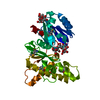
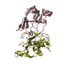
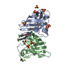
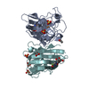
- PDB-2d6o: Crystal structure of mouse galectin-9 N-terminal CRD in complex w... -
+ Open data

- Basic information
| Entry | Database: PDB / ID: 2d6o | ||||||||||||
|---|---|---|---|---|---|---|---|---|---|---|---|---|---|
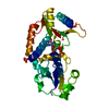
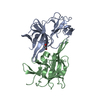
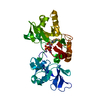
| Title | Crystal structure of mouse galectin-9 N-terminal CRD in complex with N-acetyllactosamine dimer | ||||||||||||
 Components Components | lectin, galactose binding, soluble 9 | ||||||||||||
 Keywords Keywords | SUGAR BINDING PROTEIN / beta sandwich / carbohydrate binding protein / galectin / Structural Genomics | ||||||||||||
| Function / homology |  Function and homology information Function and homology informationpositive regulation of oxidoreductase activity / negative regulation of natural killer cell degranulation / regulation of natural killer cell differentiation / negative regulation of natural killer cell activation / positive regulation of defense response to bacterium / galactoside binding / maintenance of protein location / oligosaccharide binding / positive regulation of interleukin-1 production / negative regulation of CD4-positive, alpha-beta T cell proliferation ...positive regulation of oxidoreductase activity / negative regulation of natural killer cell degranulation / regulation of natural killer cell differentiation / negative regulation of natural killer cell activation / positive regulation of defense response to bacterium / galactoside binding / maintenance of protein location / oligosaccharide binding / positive regulation of interleukin-1 production / negative regulation of CD4-positive, alpha-beta T cell proliferation / disaccharide binding / positive regulation of macrophage activation / positive regulation of innate immune response / heterophilic cell-cell adhesion / positive regulation of regulatory T cell differentiation / receptor clustering / negative regulation of type II interferon production / positive regulation of SMAD protein signal transduction / positive regulation of interleukin-10 production / positive regulation of T cell migration / immune system process / positive regulation of chemokine production / transforming growth factor beta receptor signaling pathway / positive regulation of cytokine production / protein serine/threonine kinase activator activity / female pregnancy / cellular response to virus / negative regulation of inflammatory response / positive regulation of interleukin-6 production / chemotaxis / positive regulation of tumor necrosis factor production / carbohydrate binding / extracellular matrix / response to lipopolysaccharide / receptor ligand activity / negative regulation of gene expression / signaling receptor binding / positive regulation of gene expression / enzyme binding / : / extracellular region / nucleus / plasma membrane / cytosol / cytoplasm Similarity search - Function | ||||||||||||
| Biological species |  | ||||||||||||
| Method |  X-RAY DIFFRACTION / SYNCHROTRON / MOLECULAR REPLACEMENT / Resolution: 1.78 Å X-RAY DIFFRACTION / SYNCHROTRON / MOLECULAR REPLACEMENT / Resolution: 1.78 Å | ||||||||||||
 Authors Authors | Nagae, M. / Nishi, N. / Nakamura, T. / Murata, T. / Wakatsuki, S. / Kato, R. | ||||||||||||
 Citation Citation | Journal: J.Biol.Chem. / Year: 2006 Title: Crystal Structure of the Galectin-9 N-terminal Carbohydrate Recognition Domain from Mus musculus Reveals the Basic Mechanism of Carbohydrate Recognition Authors: Nagae, M. / Nishi, N. / Murata, T. / Usui, T. / Nakamura, T. / Wakatsuki, S. / Kato, R. | ||||||||||||
| History |
|
- Structure visualization
| Structure viewer | Molecule: MolmilJmol/JSmol |
|---|
- Downloads & links
-Download
| PDBx/mmCIF format | 2d6o.cif.gz | 50.3 KB | Display | PDBx/mmCIF format |
|---|---|---|---|---|
| PDB format | pdb2d6o.ent.gz | 34.1 KB | Display | PDB format |
| PDBx/mmJSON format | 2d6o.json.gz | Tree view | PDBx/mmJSON format | |
| Others |  Other downloads Other downloads |
-Validation report
| Arichive directory | https://data.pdbj.org/pub/pdb/validation_reports/d6/2d6oftp://data.pdbj.org/pub/pdb/validation_reports/d6/2d6o | HTTPS FTP |
|---|
-Related structure data
| Related structure data | 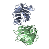 2d6kC 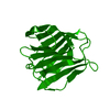 2d6lC  2d6mC 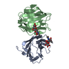 2d6nC 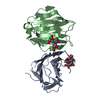 2d6pC 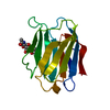 1a3kS C: citing same article ( S: Starting model for refinement |
|---|---|
| Similar structure data |

-Links
 PDBj
PDBj- Assembly
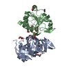
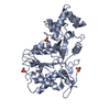
| Deposited unit | 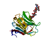
| ||||||||
|---|---|---|---|---|---|---|---|---|---|
| 1 | 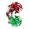
| ||||||||
| Unit cell |
|
-Components
| #1: Protein | Mass: 18169.768 Da / Num. of mol.: 1 Fragment: N-terminal carbohydrate recognition domain(RESIDUES 1-157) Source method: isolated from a genetically manipulated source Source: (gene. exp.)  |
|---|---|
| #2: Polysaccharide | beta-D-galactopyranose-(1-4)-2-acetamido-2-deoxy-beta-D-glucopyranose-(1-3)-beta-D-galactopyranose- ...beta-D-galactopyranose-(1-4)-2-acetamido-2-deoxy-beta-D-glucopyranose-(1-3)-beta-D-galactopyranose-(1-4)-2-acetamido-2-deoxy-beta-D-glucopyranose Source method: isolated from a genetically manipulated source |
| #3: Chemical | ChemComp-GOL /   Mass: 92.094 Da / Num. of mol.: 1 / Source method: obtained synthetically / Formula: C3H8O3 Mass: 92.094 Da / Num. of mol.: 1 / Source method: obtained synthetically / Formula: C3H8O3 |
| #4: Water | ChemComp-HOH /  Mass: 18.015 Da / Num. of mol.: 132 / Source method: isolated from a natural source / Formula: H2O Mass: 18.015 Da / Num. of mol.: 132 / Source method: isolated from a natural source / Formula: H2O |
| Has protein modification | Y |
-Experimental details
-Experiment
| Experiment | Method: X-RAY DIFFRACTION / Number of used crystals: 1 |
|---|
- Sample preparation
| Crystal | Density Matthews: 2.12 Å3/Da / Density % sol: 42.03 % |
|---|---|
| Crystal grow | Temperature: 289 K / Method: vapor diffusion, hanging drop / pH: 5 Details: 5% PEG6000, 0.1M citrate (pH5.0), VAPOR DIFFUSION, HANGING DROP, temperature 289K |
-Data collection
| Diffraction source | Source: SYNCHROTRON / Site: Photon Factory  / Beamline: BL-5A / Wavelength: 1 Å / Beamline: BL-5A / Wavelength: 1 Å |
|---|---|
| Detector | Type: ADSC QUANTUM 315 / Detector: CCD |
| Radiation | Protocol: SINGLE WAVELENGTH / Monochromatic (M) / Laue (L): M / Scattering type: x-ray |
| Radiation wavelength | Wavelength: 1 Å / Relative weight: 1 |
| Reflection | Resolution: 1.78→56.52 Å / Num. obs: 15123 / % possible obs: 99.8 % / Rmerge(I) obs: 0.058 |
- Processing
| Software |
| ||||||||||||||||||||||||||||||||||||||||||||||||||||||||||||||||||||||||||||||||||||||||||||||||||||||||||||||||||||||||||||||||||||||||||||||||||||||||||||||||||||||||||
|---|---|---|---|---|---|---|---|---|---|---|---|---|---|---|---|---|---|---|---|---|---|---|---|---|---|---|---|---|---|---|---|---|---|---|---|---|---|---|---|---|---|---|---|---|---|---|---|---|---|---|---|---|---|---|---|---|---|---|---|---|---|---|---|---|---|---|---|---|---|---|---|---|---|---|---|---|---|---|---|---|---|---|---|---|---|---|---|---|---|---|---|---|---|---|---|---|---|---|---|---|---|---|---|---|---|---|---|---|---|---|---|---|---|---|---|---|---|---|---|---|---|---|---|---|---|---|---|---|---|---|---|---|---|---|---|---|---|---|---|---|---|---|---|---|---|---|---|---|---|---|---|---|---|---|---|---|---|---|---|---|---|---|---|---|---|---|---|---|---|---|---|
| Refinement | Method to determine structure: MOLECULAR REPLACEMENT Starting model: PDB entry 1A3K Resolution: 1.78→56.52 Å / Cor.coef. Fo:Fc: 0.961 / Cor.coef. Fo:Fc free: 0.954 / SU B: 2.564 / SU ML: 0.082 / Cross valid method: THROUGHOUT / ESU R: 0.131 / ESU R Free: 0.12 / Stereochemistry target values: MAXIMUM LIKELIHOOD / Details: HYDROGENS HAVE BEEN ADDED IN THE RIDING POSITIONS
| ||||||||||||||||||||||||||||||||||||||||||||||||||||||||||||||||||||||||||||||||||||||||||||||||||||||||||||||||||||||||||||||||||||||||||||||||||||||||||||||||||||||||||
| Solvent computation | Ion probe radii: 0.8 Å / Shrinkage radii: 0.8 Å / VDW probe radii: 1.2 Å / Solvent model: MASK | ||||||||||||||||||||||||||||||||||||||||||||||||||||||||||||||||||||||||||||||||||||||||||||||||||||||||||||||||||||||||||||||||||||||||||||||||||||||||||||||||||||||||||
| Displacement parameters | Biso mean: 24.327 Å2
| ||||||||||||||||||||||||||||||||||||||||||||||||||||||||||||||||||||||||||||||||||||||||||||||||||||||||||||||||||||||||||||||||||||||||||||||||||||||||||||||||||||||||||
| Refinement step | Cycle: LAST / Resolution: 1.78→56.52 Å
| ||||||||||||||||||||||||||||||||||||||||||||||||||||||||||||||||||||||||||||||||||||||||||||||||||||||||||||||||||||||||||||||||||||||||||||||||||||||||||||||||||||||||||
| Refine LS restraints |
| ||||||||||||||||||||||||||||||||||||||||||||||||||||||||||||||||||||||||||||||||||||||||||||||||||||||||||||||||||||||||||||||||||||||||||||||||||||||||||||||||||||||||||
| LS refinement shell | Resolution: 1.782→1.828 Å / Total num. of bins used: 20
|
