 Movie
Movie Controller

[English] 日本語
 Yorodumi
- PDB-1a3k: X-RAY CRYSTAL STRUCTURE OF THE HUMAN GALECTIN-3 CARBOHYDRATE RECO... -
+ Open data

- Basic information
| Entry | Database: PDB / ID: 1a3k | |||||||||
|---|---|---|---|---|---|---|---|---|---|---|
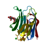
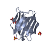
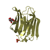
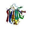
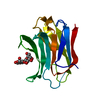
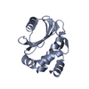
| Title | X-RAY CRYSTAL STRUCTURE OF THE HUMAN GALECTIN-3 CARBOHYDRATE RECOGNITION DOMAIN (CRD) AT 2.1 ANGSTROM RESOLUTION | |||||||||
 Components Components | GALECTIN-3 | |||||||||
 Keywords Keywords | GALECTIN / GALAPTIN / LECTIN / IGE-BINDING PROTEIN | |||||||||
| Function / homology |  Function and homology information Function and homology informationnegative regulation of NK T cell activation / negative regulation of immunological synapse formation / negative regulation of T cell activation via T cell receptor contact with antigen bound to MHC molecule on antigen presenting cell / disaccharide binding / RUNX2 regulates genes involved in differentiation of myeloid cells / regulation of T cell apoptotic process / mononuclear cell migration / receptor ligand inhibitor activity / negative regulation of endocytosis / positive regulation of mononuclear cell migration ...negative regulation of NK T cell activation / negative regulation of immunological synapse formation / negative regulation of T cell activation via T cell receptor contact with antigen bound to MHC molecule on antigen presenting cell / disaccharide binding / RUNX2 regulates genes involved in differentiation of myeloid cells / regulation of T cell apoptotic process / mononuclear cell migration / receptor ligand inhibitor activity / negative regulation of endocytosis / positive regulation of mononuclear cell migration / IgE binding / eosinophil chemotaxis / regulation of extrinsic apoptotic signaling pathway via death domain receptors / RUNX1 regulates transcription of genes involved in differentiation of myeloid cells / protein phosphatase inhibitor activity / positive chemotaxis / chemoattractant activity / positive regulation of calcium ion import / regulation of T cell proliferation / macrophage chemotaxis / Advanced glycosylation endproduct receptor signaling / monocyte chemotaxis / immunological synapse / ficolin-1-rich granule membrane / negative regulation of T cell receptor signaling pathway / neutrophil chemotaxis / epithelial cell differentiation / laminin binding / RNA splicing / secretory granule membrane / negative regulation of extrinsic apoptotic signaling pathway / positive regulation of protein localization to plasma membrane / spliceosomal complex / molecular condensate scaffold activity / positive regulation of protein-containing complex assembly / mRNA processing / carbohydrate binding / extracellular matrix / protein phosphatase binding / mitochondrial inner membrane / innate immune response / Neutrophil degranulation / cell surface / : / RNA binding / extracellular exosome / extracellular region / nucleoplasm / membrane / nucleus / plasma membrane / cytoplasm / cytosol Similarity search - Function | |||||||||
| Biological species |  Homo sapiens (human) Homo sapiens (human) | |||||||||
| Method |  X-RAY DIFFRACTION / MULTIPLE ISOMORPHOUS METHOD / Resolution: 2.1 Å X-RAY DIFFRACTION / MULTIPLE ISOMORPHOUS METHOD / Resolution: 2.1 Å | |||||||||
 Authors Authors | Seetharaman, J. / Kanigsberg, A. / Slaaby, R. / Leffler, H. / Barondes, S.H. / Rini, J.M. | |||||||||
 Citation Citation | Journal: J.Biol.Chem. / Year: 1998 Title: X-ray crystal structure of the human galectin-3 carbohydrate recognition domain at 2.1-A resolution. Authors: Seetharaman, J. / Kanigsberg, A. / Slaaby, R. / Leffler, H. / Barondes, S.H. / Rini, J.M. #1: Journal: Proc.Natl.Acad.Sci.USA / Year: 1994Title: Structure of S-Lectin, a Developmentally Regulated Vertebrate Beta-Galactoside-Binding Protein Authors: Liao, D.I. / Kapadia, G. / Ahmed, H. / Vasta, G.R. / Herzberg, O. #2: Journal: J.Biol.Chem. / Year: 1993Title: X-Ray Crystal Structure of the Human Dimeric S-Lac Lectin, L-14-II, in Complex with Lactose at 2.9-A Resolution Authors: Lobsanov, Y.D. / Gitt, M.A. / Leffler, H. / Barondes, S.H. / Rini, J.M. | |||||||||
| History |
|
- Structure visualization
| Structure viewer | Molecule: MolmilJmol/JSmol |
|---|
- Downloads & links
-Download
| PDBx/mmCIF format | 1a3k.cif.gz | 45.3 KB | Display | PDBx/mmCIF format |
|---|---|---|---|---|
| PDB format | pdb1a3k.ent.gz | 29.9 KB | Display | PDB format |
| PDBx/mmJSON format | 1a3k.json.gz | Tree view | PDBx/mmJSON format | |
| Others |  Other downloads Other downloads |
-Validation report
| Arichive directory | https://data.pdbj.org/pub/pdb/validation_reports/a3/1a3kftp://data.pdbj.org/pub/pdb/validation_reports/a3/1a3k | HTTPS FTP |
|---|
-Related structure data
| Similar structure data |
|---|


-Links
 PDBj
PDBj


- Assembly
| Deposited unit | 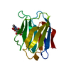
| ||||||||
|---|---|---|---|---|---|---|---|---|---|
| 1 |
| ||||||||
| Unit cell |
|
-Components
| #1: Protein | Mass: 15603.933 Da / Num. of mol.: 1 / Fragment: CARBOHYDRATE RECOGNITION DOMAIN (CRD) Source method: isolated from a genetically manipulated source Details: THE CRD WAS PRODUCED BY TYPE VII COLLAGENASE (SIGMA) DIGESTION OF THE N-TERMINAL DOMAIN Source: (gene. exp.) Homo sapiens (human)Description: THE CRD WAS PRODUCED BY TYPE VII COLLAGENASE (SIGMA) DIGESTION OF THE N-TERMINAL DOMAIN Production host:  |
|---|---|
| #2: Polysaccharide | beta-D-galactopyranose-(1-4)-2-acetamido-2-deoxy-beta-D-glucopyranose Source method: isolated from a genetically manipulated source |
| #3: Water | ChemComp-HOH /  Mass: 18.015 Da / Num. of mol.: 120 / Source method: isolated from a natural source / Formula: H2O Mass: 18.015 Da / Num. of mol.: 120 / Source method: isolated from a natural source / Formula: H2O |
-Experimental details
-Experiment
| Experiment | Method: X-RAY DIFFRACTION |
|---|
- Sample preparation
| Crystal | Density Matthews: 2.25 Å3/Da / Density % sol: 48 % | |||||||||||||||||||||||||||||||||||||||||||||||||
|---|---|---|---|---|---|---|---|---|---|---|---|---|---|---|---|---|---|---|---|---|---|---|---|---|---|---|---|---|---|---|---|---|---|---|---|---|---|---|---|---|---|---|---|---|---|---|---|---|---|---|
| Crystal grow | Method: vapor diffusion, hanging drop / pH: 8.5 Details: PROTEIN CRYSTALLIZED BY HANGING-DROP VAPOUR DIFFUSION METHOD FROM DROPS CONTAINING EQUAL VOLUMES OF PROTEIN(10-15MG/ML) IN CRYSTALLIZATION BUFFER CONTAINING 1MM LACTOSE OR 30 MM N- ...Details: PROTEIN CRYSTALLIZED BY HANGING-DROP VAPOUR DIFFUSION METHOD FROM DROPS CONTAINING EQUAL VOLUMES OF PROTEIN(10-15MG/ML) IN CRYSTALLIZATION BUFFER CONTAINING 1MM LACTOSE OR 30 MM N-ACETYLLACTOSAMINE AND THE WELL SOLUTION COMPOSED OF 27-30% PEG 4000-6000, 100MM TRIS-HCL PH 8.5, 100 MM MGCL2, AND 8 MM 2-MERCAPTOETHANOL., vapor diffusion - hanging drop | |||||||||||||||||||||||||||||||||||||||||||||||||
| Crystal grow | *PLUS Method: vapor diffusion, hanging drop | |||||||||||||||||||||||||||||||||||||||||||||||||
| Components of the solutions | *PLUS
|
-Data collection
| Diffraction | Mean temperature: 287 K |
|---|---|
| Diffraction source | Source: ROTATING ANODE / Type: OTHER / Wavelength: 1.5418 |
| Detector | Type: SIEMENS / Detector: AREA DETECTOR / Date: Dec 1, 1994 |
| Radiation | Monochromatic (M) / Laue (L): M / Scattering type: x-ray |
| Radiation wavelength | Wavelength: 1.5418 Å / Relative weight: 1 |
| Reflection | Resolution: 1.9→25 Å / Num. obs: 7955 / % possible obs: 99 % / Rmerge(I) obs: 0.052 |
| Reflection | *PLUS Num. measured all: 40093 |
- Processing
| Software |
| ||||||||||||||||||||||||||||||||||||||||||||||||||||||||||||
|---|---|---|---|---|---|---|---|---|---|---|---|---|---|---|---|---|---|---|---|---|---|---|---|---|---|---|---|---|---|---|---|---|---|---|---|---|---|---|---|---|---|---|---|---|---|---|---|---|---|---|---|---|---|---|---|---|---|---|---|---|---|
| Refinement | Method to determine structure: MULTIPLE ISOMORPHOUS METHOD / Resolution: 2.1→5 Å / σ(F): 2 Details: THE STRUCTURE WAS SOLVED BY THE MULTIPLE ISOMORPHOUS REPLACEMENT METHOD. THE MODEL CONSISTS OF THE 137 AMINO ACID RESIDUE CARBOHYDRATE RECOGNITION DOMAIN IN COMPLEX WITH N-ACETYLLACTOSAMINE ...Details: THE STRUCTURE WAS SOLVED BY THE MULTIPLE ISOMORPHOUS REPLACEMENT METHOD. THE MODEL CONSISTS OF THE 137 AMINO ACID RESIDUE CARBOHYDRATE RECOGNITION DOMAIN IN COMPLEX WITH N-ACETYLLACTOSAMINE AND 120 WATER MOLECULES. THIS STRUCTURE REPRESENTS A CRD DETERMINED FROM A GALECTIN WHICH DOES NOT SHOW THE CANONICAL 2-FOLD SYMMETRIC DIMER ORGANIZATION SHOWN BY GALECTINS-1 AND -2. COMPARISON WITH GALECTINS-1 AND -2 PROVIDES AN EXPLANATION FOR THE DIFFERENCES IN CARBOHYDRATE-BINDING SPECIFICITY SHOWN BY GALECTIN-3, AND FOR THE FACT THAT IT FAILS TO FORM DIMERS BY ANALOGOUS CRD-CRD INTERACTIONS.
| ||||||||||||||||||||||||||||||||||||||||||||||||||||||||||||
| Displacement parameters | Biso mean: 20.6 Å2 | ||||||||||||||||||||||||||||||||||||||||||||||||||||||||||||
| Refinement step | Cycle: LAST / Resolution: 2.1→5 Å
| ||||||||||||||||||||||||||||||||||||||||||||||||||||||||||||
| Refine LS restraints |
| ||||||||||||||||||||||||||||||||||||||||||||||||||||||||||||
| LS refinement shell | Resolution: 2.1→3.13 Å / Total num. of bins used: 25
| ||||||||||||||||||||||||||||||||||||||||||||||||||||||||||||
| Xplor file |
|
