 Movie
Movie Controller


+ Open data

- Basic information
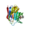
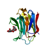
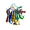
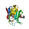
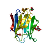
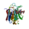
| Entry | Database: PDB / ID: 6h64 | |||||||||
|---|---|---|---|---|---|---|---|---|---|---|
| Title | Crystal structure of the CRD-SAT | |||||||||
 Components Components | Galectin-3 | |||||||||
 Keywords Keywords | SUGAR BINDING PROTEIN / carbohydrate recognition domain | |||||||||
| Function / homology |  Function and homology information Function and homology informationnegative regulation of NK T cell activation / negative regulation of immunological synapse formation / negative regulation of T cell activation via T cell receptor contact with antigen bound to MHC molecule on antigen presenting cell / disaccharide binding / RUNX2 regulates genes involved in differentiation of myeloid cells / regulation of T cell apoptotic process / mononuclear cell migration / receptor ligand inhibitor activity / negative regulation of endocytosis / positive regulation of mononuclear cell migration ...negative regulation of NK T cell activation / negative regulation of immunological synapse formation / negative regulation of T cell activation via T cell receptor contact with antigen bound to MHC molecule on antigen presenting cell / disaccharide binding / RUNX2 regulates genes involved in differentiation of myeloid cells / regulation of T cell apoptotic process / mononuclear cell migration / receptor ligand inhibitor activity / negative regulation of endocytosis / positive regulation of mononuclear cell migration / IgE binding / eosinophil chemotaxis / regulation of extrinsic apoptotic signaling pathway via death domain receptors / RUNX1 regulates transcription of genes involved in differentiation of myeloid cells / protein phosphatase inhibitor activity / positive chemotaxis / regulation of T cell proliferation / chemoattractant activity / positive regulation of calcium ion import / macrophage chemotaxis / Advanced glycosylation endproduct receptor signaling / monocyte chemotaxis / immunological synapse / ficolin-1-rich granule membrane / negative regulation of T cell receptor signaling pathway / neutrophil chemotaxis / epithelial cell differentiation / laminin binding / RNA splicing / secretory granule membrane / negative regulation of extrinsic apoptotic signaling pathway / positive regulation of protein localization to plasma membrane / spliceosomal complex / molecular condensate scaffold activity / positive regulation of protein-containing complex assembly / mRNA processing / carbohydrate binding / extracellular matrix / protein phosphatase binding / mitochondrial inner membrane / innate immune response / Neutrophil degranulation / cell surface / : / RNA binding / extracellular exosome / extracellular region / nucleoplasm / membrane / nucleus / plasma membrane / cytosol / cytoplasm Similarity search - Function | |||||||||
| Biological species |  Homo sapiens (human) Homo sapiens (human) | |||||||||
| Method |  X-RAY DIFFRACTION / SYNCHROTRON / MOLECULAR REPLACEMENT / Resolution: 1.8 Å X-RAY DIFFRACTION / SYNCHROTRON / MOLECULAR REPLACEMENT / Resolution: 1.8 Å | |||||||||
 Authors Authors | Charron, C. / Kriznik, A. / Yelehe-Okouma, M. / Jouzeau, J.-Y. / Reboul, P. | |||||||||
 Citation Citation | Journal: Biotechnol J / Year: 2019 Title: CRD SAT Generated by pCARGHO: A New Efficient Lectin-Based Affinity Tag Method for Safe, Simple, and Low-Cost Protein Purification. Authors: Kriznik, A. / Yelehe-Okouma, M. / Lec, J.C. / Groshenry, G. / Le Cordier, H. / Charron, C. / Quinternet, M. / Mazon, H. / Talfournier, F. / Boschi-Muller, S. / Jouzeau, J.Y. / Reboul, P. | |||||||||
| History |
|
- Structure visualization
| Structure viewer | Molecule: MolmilJmol/JSmol |
|---|
- Downloads & links
-Download
| PDBx/mmCIF format | 6h64.cif.gz | 192.2 KB | Display | PDBx/mmCIF format |
|---|---|---|---|---|
| PDB format | pdb6h64.ent.gz | 151.9 KB | Display | PDB format |
| PDBx/mmJSON format | 6h64.json.gz | Tree view | PDBx/mmJSON format | |
| Others |  Other downloads Other downloads |
-Validation report
| Arichive directory | https://data.pdbj.org/pub/pdb/validation_reports/h6/6h64ftp://data.pdbj.org/pub/pdb/validation_reports/h6/6h64 | HTTPS FTP |
|---|
-Related structure data
| Related structure data |  4r9bS S: Starting model for refinement |
|---|---|
| Similar structure data |


-Links
 PDBj
PDBj


- Assembly
| Deposited unit | 
| |||||||||
|---|---|---|---|---|---|---|---|---|---|---|
| 1 | 
| |||||||||
| 2 | 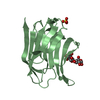
| |||||||||
| 3 | 
| |||||||||
| 4 | 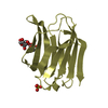
| |||||||||
| 5 | 
| |||||||||
| 6 | 
| |||||||||
| Unit cell |
| |||||||||
| Components on special symmetry positions |
|
-Components
| #1: Protein | Mass: 18836.426 Da / Num. of mol.: 6 Source method: isolated from a genetically manipulated source Source: (gene. exp.) Homo sapiens (human) / Gene: LGALS3, MAC2 / Production host:  #2: Polysaccharide | beta-D-galactopyranose-(1-4)-alpha-D-glucopyranose / alpha-lactose   Source method: isolated from a genetically manipulated source Details: oligosaccharide / References: alpha-lactose #3: Chemical | ChemComp-SO4 /   Mass: 96.063 Da / Num. of mol.: 9 / Source method: isolated from a natural source / Formula: SO4 Mass: 96.063 Da / Num. of mol.: 9 / Source method: isolated from a natural source / Formula: SO4#4: Water | ChemComp-HOH / |  Mass: 18.015 Da / Num. of mol.: 457 / Source method: isolated from a natural source / Formula: H2O Mass: 18.015 Da / Num. of mol.: 457 / Source method: isolated from a natural source / Formula: H2O |
|---|
-Experimental details
-Experiment
| Experiment | Method: X-RAY DIFFRACTION / Number of used crystals: 1 |
|---|
- Sample preparation
| Crystal | Density Matthews: 2.8 Å3/Da / Density % sol: 56.07 % |
|---|---|
| Crystal grow | Temperature: 293 K / Method: vapor diffusion, sitting drop Details: 1.6M magnesium sulfate, 100 mM MES at pH 6.5, 2 mM lactose |
-Data collection
| Diffraction | Mean temperature: 100 K |
|---|---|
| Diffraction source | Source: SYNCHROTRON / Site: SOLEIL  / Beamline: PROXIMA 1 / Wavelength: 0.979 Å / Beamline: PROXIMA 1 / Wavelength: 0.979 Å |
| Detector | Type: DECTRIS PILATUS3 6M / Detector: PIXEL / Date: Jul 7, 2016 |
| Radiation | Protocol: SINGLE WAVELENGTH / Monochromatic (M) / Laue (L): M / Scattering type: x-ray |
| Radiation wavelength | Wavelength: 0.979 Å / Relative weight: 1 |
| Reflection | Resolution: 1.8→50 Å / Num. obs: 98662 / % possible obs: 99.9 % / Redundancy: 10.3 % / Rsym value: 0.049 / Net I/σ(I): 33.3 |
| Reflection shell | Resolution: 1.8→1.91 Å |
- Processing
| Software |
| ||||||||||||||||
|---|---|---|---|---|---|---|---|---|---|---|---|---|---|---|---|---|---|
| Refinement | Method to determine structure: MOLECULAR REPLACEMENT Starting model: 4R9B Resolution: 1.8→10 Å / Cross valid method: FREE R-VALUE
| ||||||||||||||||
| Refinement step | Cycle: LAST / Resolution: 1.8→10 Å
|
