 Movie
Movie Controller

[English] 日本語
 Yorodumi
- PDB-5f12: WrbA in complex with FMN under crystallization conditions of WrbA... -
+ Open data

- Basic information
| Entry | Database: PDB / ID: 5f12 | ||||||
|---|---|---|---|---|---|---|---|
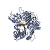
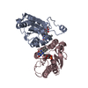
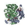
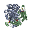
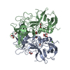
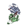
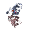
| Title | WrbA in complex with FMN under crystallization conditions of WrbA-FMN-BQ structure (4YQE) | ||||||
 Components Components | NAD(P)H dehydrogenase (quinone) | ||||||
 Keywords Keywords | OXIDOREDUCTASE / Flavin Mononucleotide / NAD(P)H Dehydrogenase (Quinone) / Oxidation-Reduction / Protein Binding / Repressor Proteins | ||||||
| Function / homology |  Function and homology information Function and homology informationNAD(P)H dehydrogenase (quinone) / NADPH dehydrogenase (quinone) activity / NADH dehydrogenase (quinone) (non-electrogenic) activity / NAD(P)H dehydrogenase (quinone) activity / NAD binding / NADP binding / FMN binding / flavin adenine dinucleotide binding / response to oxidative stress / protein-containing complex ...NAD(P)H dehydrogenase (quinone) / NADPH dehydrogenase (quinone) activity / NADH dehydrogenase (quinone) (non-electrogenic) activity / NAD(P)H dehydrogenase (quinone) activity / NAD binding / NADP binding / FMN binding / flavin adenine dinucleotide binding / response to oxidative stress / protein-containing complex / membrane / identical protein binding / cytosol Similarity search - Function | ||||||
| Biological species |  | ||||||
| Method |  X-RAY DIFFRACTION / SYNCHROTRON / MOLECULAR REPLACEMENT / molecular replacement / Resolution: 1.5 Å X-RAY DIFFRACTION / SYNCHROTRON / MOLECULAR REPLACEMENT / molecular replacement / Resolution: 1.5 Å | ||||||
 Authors Authors | Degtjarik, O. / Brynda, J. / Ettrichova, O. / Kuta Smatanova, I. / Carey, J. / Ettrich, R. | ||||||
 Citation Citation | Journal: J.Phys.Chem.B / Year: 2016 Title: Quantum Calculations Indicate Effective Electron Transfer between FMN and Benzoquinone in a New Crystal Structure of Escherichia coli WrbA. Authors: Degtjarik, O. / Brynda, J. / Ettrichova, O. / Kuty, M. / Sinha, D. / Kuta Smatanova, I. / Carey, J. / Ettrich, R. / Reha, D. | ||||||
| History |
|
- Structure visualization
| Structure viewer | Molecule: MolmilJmol/JSmol |
|---|
- Downloads & links
-Download
| PDBx/mmCIF format | 5f12.cif.gz | 91.2 KB | Display | PDBx/mmCIF format |
|---|---|---|---|---|
| PDB format | pdb5f12.ent.gz | 68.1 KB | Display | PDB format |
| PDBx/mmJSON format | 5f12.json.gz | Tree view | PDBx/mmJSON format | |
| Others |  Other downloads Other downloads |
-Validation report
| Arichive directory | https://data.pdbj.org/pub/pdb/validation_reports/f1/5f12ftp://data.pdbj.org/pub/pdb/validation_reports/f1/5f12 | HTTPS FTP |
|---|
-Related structure data
| Related structure data |  4yqeC  3b6iS C: citing same article ( S: Starting model for refinement |
|---|---|
| Similar structure data |


-Links
 PDBj
PDBj

- Assembly
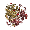
| Deposited unit | 
| ||||||||||||||||||
|---|---|---|---|---|---|---|---|---|---|---|---|---|---|---|---|---|---|---|---|
| 1 | 
| ||||||||||||||||||
| Unit cell |
| ||||||||||||||||||
| Symmetry | Point symmetry: (Schoenflies symbol: D2 (2x2 fold dihedral)) | ||||||||||||||||||
| Noncrystallographic symmetry (NCS) | NCS domain:
NCS domain segments: Component-ID: _ / Ens-ID: 1 / Beg auth comp-ID: ALA / Beg label comp-ID: ALA / End auth comp-ID: GLY / End label comp-ID: GLY / Refine code: _ / Auth seq-ID: 1 - 197 / Label seq-ID: 1 - 197
|
-Components
| #1: Protein | Mass: 20747.277 Da / Num. of mol.: 2 Source method: isolated from a genetically manipulated source Source: (gene. exp.) Gene: wrbA, b1004, JW0989 / Production host: References: UniProt: P0A8G6, NAD(P)H dehydrogenase (quinone) #2: Chemical |   Mass: 456.344 Da / Num. of mol.: 2 / Source method: obtained synthetically / Formula: C17H21N4O9P Mass: 456.344 Da / Num. of mol.: 2 / Source method: obtained synthetically / Formula: C17H21N4O9P#3: Water | ChemComp-HOH / |  Mass: 18.015 Da / Num. of mol.: 206 / Source method: isolated from a natural source / Formula: H2O Mass: 18.015 Da / Num. of mol.: 206 / Source method: isolated from a natural source / Formula: H2OHas protein modification | Y | |
|---|
-Experimental details
-Experiment
| Experiment | Method: X-RAY DIFFRACTION / Number of used crystals: 1 |
|---|
- Sample preparation
| Crystal | Density Matthews: 1.88 Å3/Da / Density % sol: 34.54 % |
|---|---|
| Crystal grow | Temperature: 295 K / Method: vapor diffusion, sitting drop / pH: 6.5 Details: 0.1 M MES/Imidazol, 12.5 % PEG 1000, 12.5% PEG 3350, 12.5% MPD, 0.2M 1,6-hexanediol, 0.2 M 1-butanol, 0.2 M (RS)-1,2-propanediol, 0.2 M 2-propanol, 0.2 M 1,4-butanediol, 0.2 M 1,3-propanediol |
-Data collection
| Diffraction | Mean temperature: 100 K |
|---|---|
| Diffraction source | Source: SYNCHROTRON / Site: BESSY  / Beamline: 14.1 / Wavelength: 0.918409 Å / Beamline: 14.1 / Wavelength: 0.918409 Å |
| Detector | Type: DECTRIS PILATUS 6M / Detector: PIXEL / Date: Sep 12, 2015 |
| Radiation | Protocol: SINGLE WAVELENGTH / Monochromatic (M) / Laue (L): M / Scattering type: x-ray |
| Radiation wavelength | Wavelength: 0.918409 Å / Relative weight: 1 |
| Reflection | Resolution: 1.5→50 Å / Num. all: 51772 / Num. obs: 51696 / % possible obs: 99.9 % / Redundancy: 8.4 % / Rmerge(I) obs: 0.1 / Net I/σ(I): 11.7 |
| Reflection shell | Resolution: 1.5→1.59 Å / Redundancy: 8.5 % / Rmerge(I) obs: 0.79 / Mean I/σ(I) obs: 2.21 / % possible all: 99.4 |
-Phasing
| Phasing | Method: molecular replacement |
|---|
- Processing
| Software |
| ||||||||||||||||||||||||||||||||||||||||||||||||||||||||||||
|---|---|---|---|---|---|---|---|---|---|---|---|---|---|---|---|---|---|---|---|---|---|---|---|---|---|---|---|---|---|---|---|---|---|---|---|---|---|---|---|---|---|---|---|---|---|---|---|---|---|---|---|---|---|---|---|---|---|---|---|---|---|
| Refinement | Method to determine structure: MOLECULAR REPLACEMENT Starting model: 3b6i Resolution: 1.5→42.96 Å / Cor.coef. Fo:Fc: 0.963 / Cor.coef. Fo:Fc free: 0.958 / WRfactor Rfree: 0.2037 / WRfactor Rwork: 0.173 / FOM work R set: 0.8566 / SU B: 1.626 / SU ML: 0.059 / SU R Cruickshank DPI: 0.0761 / SU Rfree: 0.079 / Cross valid method: THROUGHOUT / σ(F): 0 / ESU R: 0.076 / ESU R Free: 0.079 / Stereochemistry target values: MAXIMUM LIKELIHOOD Details: HYDROGENS HAVE BEEN ADDED IN THE RIDING POSITIONS U VALUES : REFINED INDIVIDUALLY
| ||||||||||||||||||||||||||||||||||||||||||||||||||||||||||||
| Solvent computation | Ion probe radii: 0.8 Å / Shrinkage radii: 0.8 Å / VDW probe radii: 1.2 Å / Solvent model: MASK | ||||||||||||||||||||||||||||||||||||||||||||||||||||||||||||
| Displacement parameters | Biso max: 79.28 Å2 / Biso mean: 21.533 Å2 / Biso min: 10.16 Å2
| ||||||||||||||||||||||||||||||||||||||||||||||||||||||||||||
| Refinement step | Cycle: final / Resolution: 1.5→42.96 Å
| ||||||||||||||||||||||||||||||||||||||||||||||||||||||||||||
| Refine LS restraints |
| ||||||||||||||||||||||||||||||||||||||||||||||||||||||||||||
| Refine LS restraints NCS | Ens-ID: 1 / Number: 10243 / Refine-ID: X-RAY DIFFRACTION / Type: interatomic distance / Rms dev position: 0.12 Å / Weight position: 0.05
| ||||||||||||||||||||||||||||||||||||||||||||||||||||||||||||
| LS refinement shell | Resolution: 1.5→1.539 Å / Total num. of bins used: 20
|
