 Movie
Movie Controller

[English] 日本語
 Yorodumi
- PDB-2ocx: Crystal structure of Se-Met fucosyltransferase NodZ from Bradyrhi... -
+ Open data

- Basic information
| Entry | Database: PDB / ID: 2ocx | ||||||
|---|---|---|---|---|---|---|---|
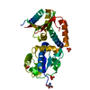
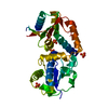
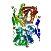
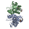
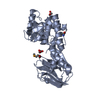
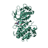
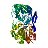
| Title | Crystal structure of Se-Met fucosyltransferase NodZ from Bradyrhizobium | ||||||
 Components Components | Nodulation fucosyltransferase NodZ | ||||||
 Keywords Keywords | TRANSFERASE / glycosyltransferase / fucosyltransferase / NodZ / nodulation | ||||||
| Function / homology |  Function and homology information Function and homology information | ||||||
| Biological species |  Bradyrhizobium sp. (bacteria) Bradyrhizobium sp. (bacteria) | ||||||
| Method |  X-RAY DIFFRACTION / SYNCHROTRON / MAD / Resolution: 2.2 Å X-RAY DIFFRACTION / SYNCHROTRON / MAD / Resolution: 2.2 Å | ||||||
 Authors Authors | Brzezinski, K. / Stepkowski, T. / Panjikar, S. / Bujacz, G. / Jaskolski, M. | ||||||
 Citation Citation | Journal: Acta Biochim.Pol. / Year: 2007 Title: High-resolution structure of NodZ fucosyltransferase involved in the biosynthesis of the nodulation factor. Authors: Brzezinski, K. / Stepkowski, T. / Panjikar, S. / Bujacz, G. / Jaskolski, M. #1: Journal: Acta Crystallogr.,Sect.D / Year: 2004Title: Cloning, purification, crystallization and preliminary crystallographic studies of Bradyrhizobium fucosyltransferase NodZ Authors: Brzezinski, K. / Rogozinski, B. / Stepkowski, T. / Bujacz, G. / Jaskolski, M. #2: Journal: J.Bacteriol. / Year: 1997Title: Rhizobium sp. strain NGR234 NodZ protein is a fucosyltransferase Authors: Quesada-Vincens, D. / Fellay, R. / Nasim, T. / Viprey, V. / Burger, U. / Prome, J.C. / Broughton, W.J. / Jabbouri, S. #3: Journal: Proc.Natl.Acad.Sci.USA / Year: 1997Title: Bacterial nodulation protein NodZ is a chitin oligosaccharide fucosyltransferase which can also recognize related substrates of animal origin Authors: Quinto, C. / Wijfjes, A.H. / Bloemberg, G.V. / Blok-Tip, L. / Lopez-Lara, I.M. / Lugtenberg, B.J. / Thomas-Oates, J.E. / Spaink, H.P. | ||||||
| History |
|
- Structure visualization
| Structure viewer | Molecule: MolmilJmol/JSmol |
|---|
- Downloads & links
-Download
| PDBx/mmCIF format | 2ocx.cif.gz | 81 KB | Display | PDBx/mmCIF format |
|---|---|---|---|---|
| PDB format | pdb2ocx.ent.gz | 60.2 KB | Display | PDB format |
| PDBx/mmJSON format | 2ocx.json.gz | Tree view | PDBx/mmJSON format | |
| Others |  Other downloads Other downloads |
-Validation report
| Arichive directory | https://data.pdbj.org/pub/pdb/validation_reports/oc/2ocxftp://data.pdbj.org/pub/pdb/validation_reports/oc/2ocx | HTTPS FTP |
|---|
-Related structure data

-Links
 PDBj
PDBj- Assembly
| Deposited unit | 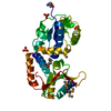
| |||||||||
|---|---|---|---|---|---|---|---|---|---|---|
| 1 |
| |||||||||
| Unit cell |
| |||||||||
| Components on special symmetry positions |
|
-Components
| #1: Protein | Mass: 38157.250 Da / Num. of mol.: 1 Source method: isolated from a genetically manipulated source Source: (gene. exp.) Bradyrhizobium sp. (bacteria) / Strain: WM9 / Gene: nodZ / Plasmid: pET3a / Production host: References: UniProt: Q9AQ17, Transferases; Glycosyltransferases; Hexosyltransferases | ||||||
|---|---|---|---|---|---|---|---|
| #2: Chemical |   Mass: 94.971 Da / Num. of mol.: 3 / Source method: obtained synthetically / Formula: PO4 Mass: 94.971 Da / Num. of mol.: 3 / Source method: obtained synthetically / Formula: PO4#3: Chemical | ChemComp-TRS / |   Mass: 122.143 Da / Num. of mol.: 1 / Source method: obtained synthetically / Formula: C4H12NO3 / Comment: pH buffer*YM Mass: 122.143 Da / Num. of mol.: 1 / Source method: obtained synthetically / Formula: C4H12NO3 / Comment: pH buffer*YM#4: Water | ChemComp-HOH / |  Mass: 18.015 Da / Num. of mol.: 180 / Source method: isolated from a natural source / Formula: H2O Mass: 18.015 Da / Num. of mol.: 180 / Source method: isolated from a natural source / Formula: H2OHas protein modification | Y | |
-Experimental details
-Experiment
| Experiment | Method: X-RAY DIFFRACTION / Number of used crystals: 1 |
|---|
- Sample preparation
| Crystal | Density Matthews: 2.82 Å3/Da / Density % sol: 56.44 % |
|---|---|
| Crystal grow | Temperature: 292 K / Method: vapor diffusion, hanging drop / pH: 7.5 Details: 0.3M potassium dihydrogen phosphate, 0.1M Tris-HCl, pH 7.5, VAPOR DIFFUSION, HANGING DROP, temperature 292K |
-Data collection
| Diffraction | Mean temperature: 100 K | ||||||||||||
|---|---|---|---|---|---|---|---|---|---|---|---|---|---|
| Diffraction source | Source: SYNCHROTRON / Site: EMBL/DESY, HAMBURG  / Beamline: BW7A / Wavelength: 0.9537, 0.9787, 0.9790 / Beamline: BW7A / Wavelength: 0.9537, 0.9787, 0.9790 | ||||||||||||
| Detector | Type: MAR CCD 165 mm / Detector: CCD / Date: Nov 20, 2004 | ||||||||||||
| Radiation | Monochromator: Si double crystal / Protocol: MAD / Monochromatic (M) / Laue (L): M / Scattering type: x-ray | ||||||||||||
| Radiation wavelength |
| ||||||||||||
| Reflection | Resolution: 2.2→20 Å / Num. all: 22641 / Num. obs: 22609 / % possible obs: 100 % / Observed criterion σ(I): -3 / Redundancy: 42.6 % / Biso Wilson estimate: 35.4 Å2 / Rmerge(I) obs: 0.085 / Net I/σ(I): 56 | ||||||||||||
| Reflection shell | Resolution: 2.2→2.28 Å / Redundancy: 43.1 % / Rmerge(I) obs: 0.346 / Mean I/σ(I) obs: 13.4 / Num. unique all: 2202 / % possible all: 100 |
- Processing
| Software |
| ||||||||||||||||||||||||||||||||||||||||||||||||||||||||||||||||||||||||||||||||||||||||||
|---|---|---|---|---|---|---|---|---|---|---|---|---|---|---|---|---|---|---|---|---|---|---|---|---|---|---|---|---|---|---|---|---|---|---|---|---|---|---|---|---|---|---|---|---|---|---|---|---|---|---|---|---|---|---|---|---|---|---|---|---|---|---|---|---|---|---|---|---|---|---|---|---|---|---|---|---|---|---|---|---|---|---|---|---|---|---|---|---|---|---|---|
| Refinement | Method to determine structure: MAD / Resolution: 2.2→19.96 Å / Cor.coef. Fo:Fc: 0.953 / Cor.coef. Fo:Fc free: 0.938 / SU B: 8.947 / SU ML: 0.12 / TLS residual ADP flag: LIKELY RESIDUAL / Cross valid method: THROUGHOUT / σ(F): 0 / ESU R: 0.197 / ESU R Free: 0.168 / Stereochemistry target values: MAXIMUM LIKELIHOOD Details: The refinement included TLS parameters. Residues 179-190, 245-256 and 318-330 were not modeled due to lack of electron density. There was no observed electron density for side chain atoms of ...Details: The refinement included TLS parameters. Residues 179-190, 245-256 and 318-330 were not modeled due to lack of electron density. There was no observed electron density for side chain atoms of residues 87, 102, 118, 119, 177, 178, 191, 193, 227, 258, 259, 305, 306, 307, 310. Those atoms were modeled with zero occupancy and B-factor of 70 A2.
| ||||||||||||||||||||||||||||||||||||||||||||||||||||||||||||||||||||||||||||||||||||||||||
| Solvent computation | Ion probe radii: 0.8 Å / Shrinkage radii: 0.8 Å / VDW probe radii: 1.4 Å / Solvent model: BABINET MODEL WITH MASK | ||||||||||||||||||||||||||||||||||||||||||||||||||||||||||||||||||||||||||||||||||||||||||
| Displacement parameters | Biso mean: 32.68 Å2
| ||||||||||||||||||||||||||||||||||||||||||||||||||||||||||||||||||||||||||||||||||||||||||
| Refinement step | Cycle: LAST / Resolution: 2.2→19.96 Å
| ||||||||||||||||||||||||||||||||||||||||||||||||||||||||||||||||||||||||||||||||||||||||||
| Refine LS restraints |
| ||||||||||||||||||||||||||||||||||||||||||||||||||||||||||||||||||||||||||||||||||||||||||
| LS refinement shell | Resolution: 2.2→2.26 Å / Total num. of bins used: 20
| ||||||||||||||||||||||||||||||||||||||||||||||||||||||||||||||||||||||||||||||||||||||||||
| Refinement TLS params. | Method: refined / Refine-ID: X-RAY DIFFRACTION
| ||||||||||||||||||||||||||||||||||||||||||||||||||||||||||||||||||||||||||||||||||||||||||
| Refinement TLS group | Refine-ID: X-RAY DIFFRACTION / Selection: ALL / Auth asym-ID: A / Label asym-ID: A
|
