 Movie
Movie Controller

[English] 日本語
 Yorodumi
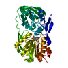
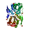
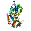
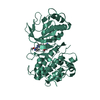
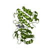
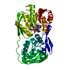
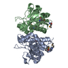
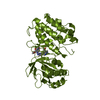
- PDB-3dmg: T. Thermophilus 16S rRNA N2 G1207 methyltransferase (RsmC) in com... -
+ Open data

- Basic information
| Entry | Database: PDB / ID: 3dmg | ||||||
|---|---|---|---|---|---|---|---|
| Title | T. Thermophilus 16S rRNA N2 G1207 methyltransferase (RsmC) in complex with AdoHcy | ||||||
 Components Components | Probable ribosomal RNA small subunit methyltransferase | ||||||
 Keywords Keywords | TRANSFERASE / monomethyltranserase / 16S rRNA methyltransferase / N2 G1207 methyltransferase / S-Adenosyl-L-Homocysteine / Translation / Methyltransferase | ||||||
| Function / homology |  Function and homology information Function and homology informationrRNA (adenine-N6,N6-)-dimethyltransferase activity / nucleic acid binding Similarity search - Function | ||||||
| Biological species |   Thermus thermophilus (bacteria) Thermus thermophilus (bacteria) | ||||||
| Method |  X-RAY DIFFRACTION / SYNCHROTRON / SAD / Resolution: 1.55 Å X-RAY DIFFRACTION / SYNCHROTRON / SAD / Resolution: 1.55 Å | ||||||
 Authors Authors | Demirci, H. / Gregory, S.T. / Dahlberg, A.E. / Jogl, G. | ||||||
 Citation Citation | Journal: J.Biol.Chem. / Year: 2008 Title: Crystal Structure of the Thermus thermophilus 16 S rRNA Methyltransferase RsmC in Complex with Cofactor and Substrate Guanosine. Authors: Demirci, H. / Gregory, S.T. / Dahlberg, A.E. / Jogl, G. | ||||||
| History |
|
- Structure visualization
| Structure viewer | Molecule: MolmilJmol/JSmol |
|---|
- Downloads & links
-Download
| PDBx/mmCIF format | 3dmg.cif.gz | 104.6 KB | Display | PDBx/mmCIF format |
|---|---|---|---|---|
| PDB format | pdb3dmg.ent.gz | 78.5 KB | Display | PDB format |
| PDBx/mmJSON format | 3dmg.json.gz | Tree view | PDBx/mmJSON format | |
| Others |  Other downloads Other downloads |
-Validation report
| Arichive directory | https://data.pdbj.org/pub/pdb/validation_reports/dm/3dmgftp://data.pdbj.org/pub/pdb/validation_reports/dm/3dmg | HTTPS FTP |
|---|
-Related structure data


-Links
 PDBj
PDBj- Assembly
| Deposited unit | 
| ||||||||
|---|---|---|---|---|---|---|---|---|---|
| 1 |
| ||||||||
| Unit cell |
|
-Components
| #1: Protein | Mass: 41537.781 Da / Num. of mol.: 1 Source method: isolated from a genetically manipulated source Details: cytoplasm / Source: (gene. exp.) Thermus thermophilus (bacteria) / Strain: HB8 / Gene: TTHA0533 / Plasmid: pET26B / Production host: References: UniProt: Q5SKW0, Transferases; Transferring one-carbon groups; Methyltransferases | ||||
|---|---|---|---|---|---|
| #2: Chemical | ChemComp-SO4 /   Mass: 96.063 Da / Num. of mol.: 6 / Source method: obtained synthetically / Formula: SO4 Mass: 96.063 Da / Num. of mol.: 6 / Source method: obtained synthetically / Formula: SO4#3: Chemical | ChemComp-SAH / |   Type: L-peptide linking / Mass: 384.411 Da / Num. of mol.: 1 / Source method: obtained synthetically / Formula: C14H20N6O5S Type: L-peptide linking / Mass: 384.411 Da / Num. of mol.: 1 / Source method: obtained synthetically / Formula: C14H20N6O5S#4: Water | ChemComp-HOH / |  Mass: 18.015 Da / Num. of mol.: 716 / Source method: isolated from a natural source / Formula: H2O Mass: 18.015 Da / Num. of mol.: 716 / Source method: isolated from a natural source / Formula: H2O |
-Experimental details
-Experiment
| Experiment | Method: X-RAY DIFFRACTION / Number of used crystals: 1 |
|---|
- Sample preparation
| Crystal | Density Matthews: 2.56 Å3/Da / Density % sol: 52 % |
|---|---|
| Crystal grow | Temperature: 277 K / Method: microbatch technique under oil / pH: 7.5 Details: 0.1 M HEPES (pH 7.5) 1.5 M lithium sulfate monohydrate, microbatch technique under oil, temperature 277K |
-Data collection
| Diffraction | Mean temperature: 100 K |
|---|---|
| Diffraction source | Source: SYNCHROTRON / Site: NSLS  / Beamline: X4C / Wavelength: 0.9797 Å / Beamline: X4C / Wavelength: 0.9797 Å |
| Detector | Type: MAR CCD 165 mm / Detector: CCD / Date: 2008 Details: Slits: Variable vertical and horizontal slits. Monochromator: Monochromator system consisting of a horizontally deflecting and focusing crystal preceded by a vertically focusing mirror. ...Details: Slits: Variable vertical and horizontal slits. Monochromator: Monochromator system consisting of a horizontally deflecting and focusing crystal preceded by a vertically focusing mirror. Distance from monochromator to sample is variable between 2.5 and 4.5 m. Distance from the monochromator to source is ~10.5 m. |
| Radiation | Monochromator: Monochromator system consisting of a horizontally deflecting and focusing crystal preceded by a vertically focusing mirror. Distance from monochromator to sample is variable between 2. ...Monochromator: Monochromator system consisting of a horizontally deflecting and focusing crystal preceded by a vertically focusing mirror. Distance from monochromator to sample is variable between 2.5 and 4.5 m. Distance from the monochromator to source is ~10.5 m. Protocol: SINGLE WAVELENGTH / Monochromatic (M) / Laue (L): M / Scattering type: x-ray |
| Radiation wavelength | Wavelength: 0.9797 Å / Relative weight: 1 |
| Reflection | Resolution: 1.55→30 Å / Num. obs: 58855 / % possible obs: 99.1 % / Redundancy: 5.5 % / Rmerge(I) obs: 0.059 / Net I/σ(I): 24.7 |
| Reflection shell | Resolution: 1.55→1.61 Å / Redundancy: 4.6 % / Rmerge(I) obs: 0.357 / Mean I/σ(I) obs: 2.6 / Num. unique all: 4186 / % possible all: 96.5 |
- Processing
| Software |
| ||||||||||||||||||||||||||||||||||||||||||||||||||||||||||||||||||||||||||||||||||||||||||||||||||||||||||||||||||||||||||||||||||||||||||||||||||||||||||||||||||||||||||
|---|---|---|---|---|---|---|---|---|---|---|---|---|---|---|---|---|---|---|---|---|---|---|---|---|---|---|---|---|---|---|---|---|---|---|---|---|---|---|---|---|---|---|---|---|---|---|---|---|---|---|---|---|---|---|---|---|---|---|---|---|---|---|---|---|---|---|---|---|---|---|---|---|---|---|---|---|---|---|---|---|---|---|---|---|---|---|---|---|---|---|---|---|---|---|---|---|---|---|---|---|---|---|---|---|---|---|---|---|---|---|---|---|---|---|---|---|---|---|---|---|---|---|---|---|---|---|---|---|---|---|---|---|---|---|---|---|---|---|---|---|---|---|---|---|---|---|---|---|---|---|---|---|---|---|---|---|---|---|---|---|---|---|---|---|---|---|---|---|---|---|---|
| Refinement | Method to determine structure: SAD / Resolution: 1.55→30 Å / Cor.coef. Fo:Fc: 0.964 / Cor.coef. Fo:Fc free: 0.95 / SU B: 2.607 / SU ML: 0.053 / TLS residual ADP flag: LIKELY RESIDUAL / Cross valid method: THROUGHOUT / ESU R: 0.084 / ESU R Free: 0.087 / Stereochemistry target values: MAXIMUM LIKELIHOOD Details: HYDROGENS HAVE BEEN ADDED IN THE RIDING POSITIONS. FOR STRUCTURE DETERMINATION, AUTHORS STATE THAT THEY SOLVED THESE STRUCTURES BY USING AN ANOMALOUS DATA FROM ANOTHER CRYSTAL. AUTHORS ...Details: HYDROGENS HAVE BEEN ADDED IN THE RIDING POSITIONS. FOR STRUCTURE DETERMINATION, AUTHORS STATE THAT THEY SOLVED THESE STRUCTURES BY USING AN ANOMALOUS DATA FROM ANOTHER CRYSTAL. AUTHORS REFINED THESE DATASETS BY USING PREVIOUS MODEL WITHOUT ANY MOLECULAR REPLACEMENT.
| ||||||||||||||||||||||||||||||||||||||||||||||||||||||||||||||||||||||||||||||||||||||||||||||||||||||||||||||||||||||||||||||||||||||||||||||||||||||||||||||||||||||||||
| Solvent computation | Ion probe radii: 0.8 Å / Shrinkage radii: 0.8 Å / VDW probe radii: 1.4 Å / Solvent model: MASK | ||||||||||||||||||||||||||||||||||||||||||||||||||||||||||||||||||||||||||||||||||||||||||||||||||||||||||||||||||||||||||||||||||||||||||||||||||||||||||||||||||||||||||
| Displacement parameters | Biso mean: 26.52 Å2
| ||||||||||||||||||||||||||||||||||||||||||||||||||||||||||||||||||||||||||||||||||||||||||||||||||||||||||||||||||||||||||||||||||||||||||||||||||||||||||||||||||||||||||
| Refinement step | Cycle: LAST / Resolution: 1.55→30 Å
| ||||||||||||||||||||||||||||||||||||||||||||||||||||||||||||||||||||||||||||||||||||||||||||||||||||||||||||||||||||||||||||||||||||||||||||||||||||||||||||||||||||||||||
| Refine LS restraints |
| ||||||||||||||||||||||||||||||||||||||||||||||||||||||||||||||||||||||||||||||||||||||||||||||||||||||||||||||||||||||||||||||||||||||||||||||||||||||||||||||||||||||||||
| LS refinement shell | Resolution: 1.55→1.59 Å / Total num. of bins used: 20
| ||||||||||||||||||||||||||||||||||||||||||||||||||||||||||||||||||||||||||||||||||||||||||||||||||||||||||||||||||||||||||||||||||||||||||||||||||||||||||||||||||||||||||
| Refinement TLS params. | Method: refined / Refine-ID: X-RAY DIFFRACTION
| ||||||||||||||||||||||||||||||||||||||||||||||||||||||||||||||||||||||||||||||||||||||||||||||||||||||||||||||||||||||||||||||||||||||||||||||||||||||||||||||||||||||||||
| Refinement TLS group |
|
