 Movie
Movie Controller

[English] 日本語
 Yorodumi
- PDB-3s0q: Peptidase module of the peptidoglycan hydrolase RipA (Rv1477) fro... -
+ Open data

- Basic information
| Entry | Database: PDB / ID: 3s0q | ||||||
|---|---|---|---|---|---|---|---|
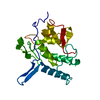
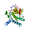
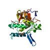
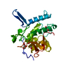
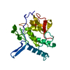
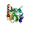
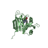
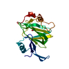
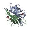
| Title | Peptidase module of the peptidoglycan hydrolase RipA (Rv1477) from Mycobacterium tuberculosis, catalytic site mutant (Cys383Ala) at 1.45 resolution | ||||||
 Components Components | INVASION PROTEIN | ||||||
 Keywords Keywords | HYDROLASE / NlpC/p60 module / peptidoglycan hydrolase | ||||||
| Function / homology |  Function and homology information Function and homology informationcell wall organization or biogenesis / N-acetylmuramoyl-L-alanine amidase activity / Hydrolases; Acting on peptide bonds (peptidases) / cysteine-type peptidase activity / peptidoglycan-based cell wall / cell wall organization / proteolysis / extracellular region Similarity search - Function | ||||||
| Biological species |   Mycobacterium tuberculosis (bacteria) Mycobacterium tuberculosis (bacteria) | ||||||
| Method |  X-RAY DIFFRACTION / SYNCHROTRON / MOLECULAR REPLACEMENT / Resolution: 1.45 Å X-RAY DIFFRACTION / SYNCHROTRON / MOLECULAR REPLACEMENT / Resolution: 1.45 Å | ||||||
 Authors Authors | Both, D. / Schnell, R. / Schneider, G. | ||||||
 Citation Citation | Journal: J.Mol.Biol. / Year: 2011 Title: Peptidoglycan Remodeling in Mycobacterium tuberculosis: Comparison of Structures and Catalytic Activities of RipA and RipB. Authors: Both, D. / Schneider, G. / Schnell, R. | ||||||
| History |
|
- Structure visualization
| Structure viewer | Molecule: MolmilJmol/JSmol |
|---|
- Downloads & links
-Download
| PDBx/mmCIF format | 3s0q.cif.gz | 56.6 KB | Display | PDBx/mmCIF format |
|---|---|---|---|---|
| PDB format | pdb3s0q.ent.gz | 40.4 KB | Display | PDB format |
| PDBx/mmJSON format | 3s0q.json.gz | Tree view | PDBx/mmJSON format | |
| Others |  Other downloads Other downloads |
-Validation report
| Arichive directory | https://data.pdbj.org/pub/pdb/validation_reports/s0/3s0qftp://data.pdbj.org/pub/pdb/validation_reports/s0/3s0q | HTTPS FTP |
|---|
-Related structure data


-Links
 PDBj
PDBj- Assembly
| Deposited unit | 
| ||||||||
|---|---|---|---|---|---|---|---|---|---|
| 1 |
| ||||||||
| Unit cell |
|
-Components
| #1: Protein | Mass: 22706.783 Da / Num. of mol.: 1 / Fragment: RipA peptidase module / Mutation: C383A Source method: isolated from a genetically manipulated source Source: (gene. exp.) Mycobacterium tuberculosis (bacteria) / Strain: H37Rv / Gene: Rv1477 / Plasmid: pNIC28Bsa4 / Production host: |
|---|---|
| #2: Water | ChemComp-HOH /  Mass: 18.015 Da / Num. of mol.: 244 / Source method: isolated from a natural source / Formula: H2O Mass: 18.015 Da / Num. of mol.: 244 / Source method: isolated from a natural source / Formula: H2O |
-Experimental details
-Experiment
| Experiment | Method: X-RAY DIFFRACTION / Number of used crystals: 1 |
|---|
- Sample preparation
| Crystal | Density Matthews: 1.82 Å3/Da / Density % sol: 32.27 % |
|---|---|
| Crystal grow | Temperature: 293 K / Method: vapor diffusion, hanging drop / pH: 8.5 Details: 35% PEG400, 0.2M LiSO4, Tris-HCl pH 8.5, VAPOR DIFFUSION, HANGING DROP, temperature 293K |
-Data collection
| Diffraction | Mean temperature: 100 K |
|---|---|
| Diffraction source | Source: SYNCHROTRON / Site: MAX II  / Beamline: I911-2 / Wavelength: 1.03873 Å / Beamline: I911-2 / Wavelength: 1.03873 Å |
| Detector | Type: MAR CCD 165 mm / Detector: CCD / Date: Feb 1, 2011 Details: Multilayer mirror, curved to focus in the vertical (R = 400 m). |
| Radiation | Monochromator: Bent Si (111) crystal, horizontally focusing / Protocol: SINGLE WAVELENGTH / Monochromatic (M) / Laue (L): M / Scattering type: x-ray |
| Radiation wavelength | Wavelength: 1.03873 Å / Relative weight: 1 |
| Reflection | Resolution: 1.45→18.67 Å / Num. all: 30245 / Num. obs: 29973 / % possible obs: 99.1 % / Observed criterion σ(F): 4 / Observed criterion σ(I): 4 / Redundancy: 4.8 % / Biso Wilson estimate: 10.7 Å2 / Rmerge(I) obs: 0.068 / Rsym value: 0.068 / Net I/σ(I): 15.1 |
| Reflection shell | Resolution: 1.45→1.53 Å / Redundancy: 4.6 % / Rmerge(I) obs: 0.375 / Mean I/σ(I) obs: 4 / Num. unique all: 4098 / Rsym value: 0.375 / % possible all: 94.4 |
- Processing
| Software |
| |||||||||||||||||||||||||||||||||||||||||||||||||||||||||||||||||||||||||||||||||||||
|---|---|---|---|---|---|---|---|---|---|---|---|---|---|---|---|---|---|---|---|---|---|---|---|---|---|---|---|---|---|---|---|---|---|---|---|---|---|---|---|---|---|---|---|---|---|---|---|---|---|---|---|---|---|---|---|---|---|---|---|---|---|---|---|---|---|---|---|---|---|---|---|---|---|---|---|---|---|---|---|---|---|---|---|---|---|---|
| Refinement | Method to determine structure: MOLECULAR REPLACEMENT / Resolution: 1.45→18.57 Å / Cor.coef. Fo:Fc: 0.966 / Cor.coef. Fo:Fc free: 0.958 / SU B: 0.889 / SU ML: 0.035 / Isotropic thermal model: isotropic / Cross valid method: THROUGHOUT / σ(F): 4 / ESU R Free: 0.067 / Stereochemistry target values: MAXIMUM LIKELIHOOD / Details: HYDROGENS HAVE BEEN ADDED IN THE RIDING POSITIONS
| |||||||||||||||||||||||||||||||||||||||||||||||||||||||||||||||||||||||||||||||||||||
| Solvent computation | Ion probe radii: 0.8 Å / Shrinkage radii: 0.8 Å / VDW probe radii: 1.4 Å / Solvent model: MASK | |||||||||||||||||||||||||||||||||||||||||||||||||||||||||||||||||||||||||||||||||||||
| Displacement parameters | Biso mean: 7.543 Å2
| |||||||||||||||||||||||||||||||||||||||||||||||||||||||||||||||||||||||||||||||||||||
| Refine analyze | Luzzati coordinate error free: 0.067 Å | |||||||||||||||||||||||||||||||||||||||||||||||||||||||||||||||||||||||||||||||||||||
| Refinement step | Cycle: LAST / Resolution: 1.45→18.57 Å
| |||||||||||||||||||||||||||||||||||||||||||||||||||||||||||||||||||||||||||||||||||||
| Refine LS restraints |
| |||||||||||||||||||||||||||||||||||||||||||||||||||||||||||||||||||||||||||||||||||||
| LS refinement shell | Resolution: 1.449→1.487 Å / Total num. of bins used: 20
|
