 Movie
Movie Controller

[English] 日本語
 Yorodumi
- PDB-3ne0: Structure and functional regulation of RipA, a mycobacterial enzy... -
+ Open data

- Basic information
| Entry | Database: PDB / ID: 3ne0 | ||||||
|---|---|---|---|---|---|---|---|
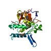
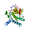
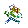
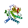
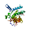
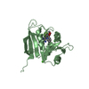
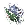
| Title | Structure and functional regulation of RipA, a mycobacterial enzyme essential for daughter cell separation | ||||||
 Components Components | Resuscitation promoting factor Interacting Protein A | ||||||
 Keywords Keywords | HYDROLASE / cell wall / peptidoglycan / tuberculosis | ||||||
| Function / homology |  Function and homology information Function and homology informationcell wall organization or biogenesis / N-acetylmuramoyl-L-alanine amidase activity / Hydrolases; Acting on peptide bonds (peptidases) / cysteine-type peptidase activity / peptidoglycan-based cell wall / cell wall organization / proteolysis / extracellular region Similarity search - Function | ||||||
| Biological species |   Mycobacterium tuberculosis (bacteria) Mycobacterium tuberculosis (bacteria) | ||||||
| Method |  X-RAY DIFFRACTION / SYNCHROTRON / MAD / Resolution: 1 Å X-RAY DIFFRACTION / SYNCHROTRON / MAD / Resolution: 1 Å | ||||||
 Authors Authors | Ruggiero, A. / Squeglia, F. / Berisio, R. | ||||||
 Citation Citation | Journal: Structure / Year: 2010 Title: Structure and Functional Regulation of RipA, a Mycobacterial Enzyme Essential for Daughter Cell Separation. Authors: Ruggiero, A. / Marasco, D. / Squeglia, F. / Soldini, S. / Pedone, E. / Pedone, C. / Berisio, R. | ||||||
| History |
|
- Structure visualization
| Structure viewer | Molecule: MolmilJmol/JSmol |
|---|
- Downloads & links
-Download
| PDBx/mmCIF format | 3ne0.cif.gz | 106.3 KB | Display | PDBx/mmCIF format |
|---|---|---|---|---|
| PDB format | pdb3ne0.ent.gz | 80.1 KB | Display | PDB format |
| PDBx/mmJSON format | 3ne0.json.gz | Tree view | PDBx/mmJSON format | |
| Others |  Other downloads Other downloads |
-Validation report
| Arichive directory | https://data.pdbj.org/pub/pdb/validation_reports/ne/3ne0ftp://data.pdbj.org/pub/pdb/validation_reports/ne/3ne0 | HTTPS FTP |
|---|
-Related structure data
| Related structure data | |
|---|---|
| Similar structure data |


-Links
 PDBj
PDBj- Assembly
| Deposited unit | 
| ||||||||
|---|---|---|---|---|---|---|---|---|---|
| 1 |
| ||||||||
| Unit cell |
|
-Components
| #1: Protein | Mass: 22551.629 Da / Num. of mol.: 1 / Fragment: UNP residues 263-472 Source method: isolated from a genetically manipulated source Source: (gene. exp.) Mycobacterium tuberculosis (bacteria) / Strain: h37rv / Gene: rv1477 / Production host: |
|---|---|
| #2: Water | ChemComp-HOH /  Mass: 18.015 Da / Num. of mol.: 390 / Source method: isolated from a natural source / Formula: H2O Mass: 18.015 Da / Num. of mol.: 390 / Source method: isolated from a natural source / Formula: H2O |
-Experimental details
-Experiment
| Experiment | Method: X-RAY DIFFRACTION / Number of used crystals: 1 |
|---|
- Sample preparation
| Crystal | Density Matthews: 1.82 Å3/Da / Density % sol: 32.42 % |
|---|---|
| Crystal grow | Temperature: 293 K / Method: evaporation / pH: 5.6 Details: 8% (v/v) 2-Propanol, 16% (w/v) PEG4000, 60 mM Sodium citrate trihydrate buffer, pH 5.6, EVAPORATION, temperature 293K |
-Data collection
| Diffraction | Mean temperature: 100 K | ||||||||||||
|---|---|---|---|---|---|---|---|---|---|---|---|---|---|
| Diffraction source | Source: SYNCHROTRON / Site: ESRF  / Beamline: BM14 / Wavelength: 0.9737, 0.9785, 0.9787, 0.9737 / Beamline: BM14 / Wavelength: 0.9737, 0.9785, 0.9787, 0.9737 | ||||||||||||
| Detector | Type: MAR scanner 345 mm plate / Detector: IMAGE PLATE / Date: Jun 30, 2008 / Details: mirrors | ||||||||||||
| Radiation | Monochromator: Si(111) monochromator. / Protocol: MAD / Monochromatic (M) / Laue (L): M / Scattering type: x-ray | ||||||||||||
| Radiation wavelength |
| ||||||||||||
| Reflection | Resolution: 1→30 Å / Num. all: 88997 / Num. obs: 85882 / % possible obs: 96.5 % / Observed criterion σ(F): 0 / Observed criterion σ(I): 0 / Rmerge(I) obs: 0.08 / Net I/σ(I): 19.7 | ||||||||||||
| Reflection shell | Resolution: 1→1.04 Å / Redundancy: 3.1 % / Rmerge(I) obs: 0.292 / Mean I/σ(I) obs: 3 / % possible all: 72.6 |
- Processing
| Software |
| |||||||||||||||||||||||||||||||||
|---|---|---|---|---|---|---|---|---|---|---|---|---|---|---|---|---|---|---|---|---|---|---|---|---|---|---|---|---|---|---|---|---|---|---|
| Refinement | Method to determine structure: MAD / Resolution: 1→10 Å / Num. parameters: 17484 / Num. restraintsaints: 7222 / σ(F): 0 / Stereochemistry target values: ENGH AND HUBER
| |||||||||||||||||||||||||||||||||
| Refine analyze | Num. disordered residues: 2 / Occupancy sum hydrogen: 1526 / Occupancy sum non hydrogen: 1934 | |||||||||||||||||||||||||||||||||
| Refinement step | Cycle: LAST / Resolution: 1→10 Å
| |||||||||||||||||||||||||||||||||
| Refine LS restraints |
| |||||||||||||||||||||||||||||||||
| LS refinement shell | Resolution: 1→1.04 Å /
|
