 Movie
Movie Controller


+ Open data

- Basic information
| Entry | Database: PDB / ID: 6l5a | ||||||
|---|---|---|---|---|---|---|---|
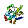
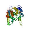
| Title | The structure of the UdgX mutant H109E at a pre-excision state | ||||||
 Components Components | Uracil DNA glycosylase superfamily protein | ||||||
 Keywords Keywords | DNA BINDING PROTEIN / UdgX / covalent complex / protein-DNA interaction / DNA repair / glycosylase | ||||||
| Function / homology |  Function and homology information Function and homology informationdeaminated base DNA N-glycosylase activity / 4 iron, 4 sulfur cluster binding / DNA repair / metal ion binding Similarity search - Function | ||||||
| Biological species |  Mycolicibacterium smegmatis MC2 155 (bacteria) Mycolicibacterium smegmatis MC2 155 (bacteria) | ||||||
| Method |  X-RAY DIFFRACTION / MOLECULAR REPLACEMENT / Resolution: 1.80007323734 Å X-RAY DIFFRACTION / MOLECULAR REPLACEMENT / Resolution: 1.80007323734 Å | ||||||
 Authors Authors | Xie, W. / Tu, J. / Zeng, H. | ||||||
| Funding support |  China, 1items China, 1items
| ||||||
 Citation Citation | Journal: DNA Repair (Amst) / Year: 2021 Title: Structural insights into an MsmUdgX mutant capable of both crosslinking and uracil excision capability. Authors: Jia, Q. / Zeng, H. / Tu, J. / Sun, L. / Cao, W. / Xie, W. | ||||||
| History |
|
- Structure visualization
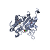
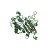
| Structure viewer | Molecule: MolmilJmol/JSmol |
|---|
- Downloads & links
-Download
| PDBx/mmCIF format | 6l5a.cif.gz | 74 KB | Display | PDBx/mmCIF format |
|---|---|---|---|---|
| PDB format | pdb6l5a.ent.gz | 42 KB | Display | PDB format |
| PDBx/mmJSON format | 6l5a.json.gz | Tree view | PDBx/mmJSON format | |
| Others |  Other downloads Other downloads |
-Validation report
| Arichive directory | https://data.pdbj.org/pub/pdb/validation_reports/l5/6l5aftp://data.pdbj.org/pub/pdb/validation_reports/l5/6l5a | HTTPS FTP |
|---|
-Related structure data
| Related structure data | 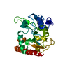 6l5bC  6l6sC  6io9S S: Starting model for refinement C: citing same article ( |
|---|---|
| Similar structure data |


-Links
 PDBj
PDBj- Assembly
| Deposited unit | 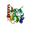
| ||||||||||||
|---|---|---|---|---|---|---|---|---|---|---|---|---|---|
| 1 |
| ||||||||||||
| Unit cell |
|
-Components
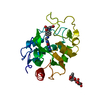
| #1: Protein | Mass: 23249.420 Da / Num. of mol.: 1 / Mutation: H109E Source method: isolated from a genetically manipulated source Source: (gene. exp.) Mycolicibacterium smegmatis MC2 155 (bacteria)Strain: MC2 155 / Gene: MSMEG_0265 / Production host: | ||||
|---|---|---|---|---|---|
| #2: Chemical | ChemComp-SF4 /   Mass: 351.640 Da / Num. of mol.: 1 / Source method: obtained synthetically / Formula: Fe4S4 / Feature type: SUBJECT OF INVESTIGATION Mass: 351.640 Da / Num. of mol.: 1 / Source method: obtained synthetically / Formula: Fe4S4 / Feature type: SUBJECT OF INVESTIGATION | ||||
| #3: Chemical |   Mass: 92.094 Da / Num. of mol.: 2 / Source method: obtained synthetically / Formula: C3H8O3 / Feature type: SUBJECT OF INVESTIGATION Mass: 92.094 Da / Num. of mol.: 2 / Source method: obtained synthetically / Formula: C3H8O3 / Feature type: SUBJECT OF INVESTIGATION#4: Water | ChemComp-HOH / |  Mass: 18.015 Da / Num. of mol.: 268 / Source method: isolated from a natural source / Formula: H2O Mass: 18.015 Da / Num. of mol.: 268 / Source method: isolated from a natural source / Formula: H2OHas ligand of interest | Y | |
-Experimental details
-Experiment
| Experiment | Method: X-RAY DIFFRACTION / Number of used crystals: 1 |
|---|
- Sample preparation
| Crystal | Density Matthews: 2.14 Å3/Da / Density % sol: 42.43 % |
|---|---|
| Crystal grow | Temperature: 298 K / Method: vapor diffusion, sitting drop / pH: 5.5 / Details: 19% PEG3350, 0.1 M sodium citrate, 0.2 M NaCl |
-Data collection
| Diffraction | Mean temperature: 100 K / Serial crystal experiment: N |
|---|---|
| Diffraction source | Source: ROTATING ANODE / Type: RIGAKU MICROMAX-007 HF / Wavelength: 1.54 Å |
| Detector | Type: OXFORD ONYX CCD / Detector: CCD / Date: Jun 11, 2019 |
| Radiation | Protocol: SINGLE WAVELENGTH / Monochromatic (M) / Laue (L): M / Scattering type: x-ray |
| Radiation wavelength | Wavelength: 1.54 Å / Relative weight: 1 |
| Reflection | Resolution: 1.8→21.56 Å / Num. obs: 18687 / % possible obs: 98.1 % / Redundancy: 4.8 % / Biso Wilson estimate: 8.78342573865 Å2 / CC1/2: 0.998 / Rmerge(I) obs: 0.064 / Net I/σ(I): 15.9 |
| Reflection shell | Resolution: 1.8→1.9 Å / Redundancy: 4.2 % / Rmerge(I) obs: 0.128 / Mean I/σ(I) obs: 8.4 / Num. unique obs: 2586 / CC1/2: 0.985 / % possible all: 95.1 |
- Processing
| Software |
| ||||||||||||||||||||||||||||||||||||||||||||||||||||||||
|---|---|---|---|---|---|---|---|---|---|---|---|---|---|---|---|---|---|---|---|---|---|---|---|---|---|---|---|---|---|---|---|---|---|---|---|---|---|---|---|---|---|---|---|---|---|---|---|---|---|---|---|---|---|---|---|---|---|
| Refinement | Method to determine structure: MOLECULAR REPLACEMENT Starting model: 6IO9 Resolution: 1.80007323734→21.55545 Å / SU ML: 0.155071477148 / Cross valid method: FREE R-VALUE / σ(F): 1.3379521463 / Phase error: 17.4976843893
| ||||||||||||||||||||||||||||||||||||||||||||||||||||||||
| Solvent computation | Shrinkage radii: 0.9 Å / VDW probe radii: 1.11 Å | ||||||||||||||||||||||||||||||||||||||||||||||||||||||||
| Displacement parameters | Biso mean: 11.6844359257 Å2 | ||||||||||||||||||||||||||||||||||||||||||||||||||||||||
| Refinement step | Cycle: LAST / Resolution: 1.80007323734→21.55545 Å
| ||||||||||||||||||||||||||||||||||||||||||||||||||||||||
| Refine LS restraints |
| ||||||||||||||||||||||||||||||||||||||||||||||||||||||||
| LS refinement shell |
|
