 Movie
Movie Controller

[English] 日本語
 Yorodumi
- PDB-3pbi: Structure of the peptidoglycan hydrolase RipB (Rv1478) from Mycob... -
+ Open data

- Basic information
| Entry | Database: PDB / ID: 3pbi | ||||||
|---|---|---|---|---|---|---|---|
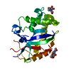
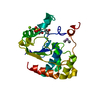
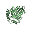
| Title | Structure of the peptidoglycan hydrolase RipB (Rv1478) from Mycobacterium tuberculosis at 1.6 resolution | ||||||
 Components Components | INVASION PROTEIN | ||||||
 Keywords Keywords | HYDROLASE / peptidoglycan hydrolase / extracellular / invasion related protein / cell wall / NlpC-like module | ||||||
| Function / homology |  Function and homology information Function and homology informationcell wall organization or biogenesis / cell wall / N-acetylmuramoyl-L-alanine amidase activity / Hydrolases; Acting on peptide bonds (peptidases) / cysteine-type peptidase activity / peptidoglycan-based cell wall / cell wall organization / proteolysis Similarity search - Function | ||||||
| Biological species |   Mycobacterium tuberculosis (bacteria) Mycobacterium tuberculosis (bacteria) | ||||||
| Method |  X-RAY DIFFRACTION / SYNCHROTRON / MOLECULAR REPLACEMENT / Resolution: 1.6 Å X-RAY DIFFRACTION / SYNCHROTRON / MOLECULAR REPLACEMENT / Resolution: 1.6 Å | ||||||
 Authors Authors | Schnell, R. / Both, D. / Schneider, G. | ||||||
 Citation Citation | Journal: J.Mol.Biol. / Year: 2011 Title: Peptidoglycan Remodeling in Mycobacterium tuberculosis: Comparison of Structures and Catalytic Activities of RipA and RipB. Authors: Both, D. / Schneider, G. / Schnell, R. | ||||||
| History |
|
- Structure visualization
| Structure viewer | Molecule: MolmilJmol/JSmol |
|---|
- Downloads & links
-Download
| PDBx/mmCIF format | 3pbi.cif.gz | 56.2 KB | Display | PDBx/mmCIF format |
|---|---|---|---|---|
| PDB format | pdb3pbi.ent.gz | 39.3 KB | Display | PDB format |
| PDBx/mmJSON format | 3pbi.json.gz | Tree view | PDBx/mmJSON format | |
| Others |  Other downloads Other downloads |
-Validation report
| Arichive directory | https://data.pdbj.org/pub/pdb/validation_reports/pb/3pbiftp://data.pdbj.org/pub/pdb/validation_reports/pb/3pbi | HTTPS FTP |
|---|
-Related structure data
| Related structure data |  3pbcC 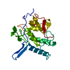 3s0qC  2hbwS C: citing same article ( S: Starting model for refinement |
|---|---|
| Similar structure data |


-Links
 PDBj
PDBj- Assembly
| Deposited unit | 
| ||||||||
|---|---|---|---|---|---|---|---|---|---|
| 1 |
| ||||||||
| Unit cell |
|
-Components
| #1: Protein | Mass: 21733.350 Da / Num. of mol.: 1 / Fragment: unp residues 30-241 Source method: isolated from a genetically manipulated source Source: (gene. exp.) Mycobacterium tuberculosis (bacteria) / Strain: H37Rv / Gene: MT1525, Rv1478 / Plasmid: pNIC28Bsa4 / Production host: |
|---|---|
| #2: Water | ChemComp-HOH /  Mass: 18.015 Da / Num. of mol.: 242 / Source method: isolated from a natural source / Formula: H2O Mass: 18.015 Da / Num. of mol.: 242 / Source method: isolated from a natural source / Formula: H2O |
-Experimental details
-Experiment
| Experiment | Method: X-RAY DIFFRACTION / Number of used crystals: 1 |
|---|
- Sample preparation
| Crystal | Density Matthews: 3.07 Å3/Da / Density % sol: 59.97 % |
|---|---|
| Crystal grow | Temperature: 293 K / pH: 7.5 Details: 0.2 M NaCl 0.1 M HEPES, 30% v/v PEG 400, pH 7.5, VAPOR DIFFUSION, SITTING DROP, temperature 293K |
-Data collection
| Diffraction | Mean temperature: 100 K |
|---|---|
| Diffraction source | Source: SYNCHROTRON / Site: ESRF  / Beamline: ID23-1 / Wavelength: 0.9794 / Beamline: ID23-1 / Wavelength: 0.9794 |
| Detector | Type: ADSC QUANTUM 315r / Detector: CCD / Date: Oct 25, 2009 / Details: MIRRORS |
| Radiation | Monochromator: SI (311) MONOCHROMATOR / Protocol: SINGLE WAVELENGTH / Monochromatic (M) / Laue (L): M / Scattering type: x-ray |
| Radiation wavelength | Wavelength: 0.9794 Å / Relative weight: 1 |
| Reflection | Resolution: 1.6→40 Å / Num. obs: 32063 / % possible obs: 92.3 % / Observed criterion σ(I): 2.5 / Redundancy: 7.4 % / Biso Wilson estimate: 20.1 Å2 / Rmerge(I) obs: 0.066 / Rsym value: 0.066 / Net I/σ(I): 19.1 |
| Reflection shell | Resolution: 1.6→1.69 Å / Redundancy: 2.8 % / Rmerge(I) obs: 0.411 / Mean I/σ(I) obs: 2.5 / Rsym value: 0.411 / % possible all: 63.2 |
- Processing
| Software |
| ||||||||||||||||||||||||||||||||||||||||||||||||||||||||||||||||||||||||||||||||||||||||||||||||||||||||||||||||||||||||||||||||||||||||||||||||||||||||||||||||||||||||||
|---|---|---|---|---|---|---|---|---|---|---|---|---|---|---|---|---|---|---|---|---|---|---|---|---|---|---|---|---|---|---|---|---|---|---|---|---|---|---|---|---|---|---|---|---|---|---|---|---|---|---|---|---|---|---|---|---|---|---|---|---|---|---|---|---|---|---|---|---|---|---|---|---|---|---|---|---|---|---|---|---|---|---|---|---|---|---|---|---|---|---|---|---|---|---|---|---|---|---|---|---|---|---|---|---|---|---|---|---|---|---|---|---|---|---|---|---|---|---|---|---|---|---|---|---|---|---|---|---|---|---|---|---|---|---|---|---|---|---|---|---|---|---|---|---|---|---|---|---|---|---|---|---|---|---|---|---|---|---|---|---|---|---|---|---|---|---|---|---|---|---|---|
| Refinement | Method to determine structure: MOLECULAR REPLACEMENT Starting model: PDB ENTRY 2HBW Resolution: 1.6→40 Å / Cor.coef. Fo:Fc: 0.967 / Cor.coef. Fo:Fc free: 0.948 / SU B: 1.332 / SU ML: 0.047 / Cross valid method: THROUGHOUT / σ(F): 2.5 / ESU R Free: 0.078 / Stereochemistry target values: MAXIMUM LIKELIHOOD / Details: HYDROGENS HAVE BEEN ADDED IN THE RIDING POSITIONS
| ||||||||||||||||||||||||||||||||||||||||||||||||||||||||||||||||||||||||||||||||||||||||||||||||||||||||||||||||||||||||||||||||||||||||||||||||||||||||||||||||||||||||||
| Solvent computation | Ion probe radii: 0.8 Å / Shrinkage radii: 0.8 Å / VDW probe radii: 1.4 Å / Solvent model: BABINET MODEL WITH MASK | ||||||||||||||||||||||||||||||||||||||||||||||||||||||||||||||||||||||||||||||||||||||||||||||||||||||||||||||||||||||||||||||||||||||||||||||||||||||||||||||||||||||||||
| Displacement parameters | Biso mean: 19.26 Å2
| ||||||||||||||||||||||||||||||||||||||||||||||||||||||||||||||||||||||||||||||||||||||||||||||||||||||||||||||||||||||||||||||||||||||||||||||||||||||||||||||||||||||||||
| Refine analyze |
| ||||||||||||||||||||||||||||||||||||||||||||||||||||||||||||||||||||||||||||||||||||||||||||||||||||||||||||||||||||||||||||||||||||||||||||||||||||||||||||||||||||||||||
| Refinement step | Cycle: LAST / Resolution: 1.6→40 Å
| ||||||||||||||||||||||||||||||||||||||||||||||||||||||||||||||||||||||||||||||||||||||||||||||||||||||||||||||||||||||||||||||||||||||||||||||||||||||||||||||||||||||||||
| Refine LS restraints |
| ||||||||||||||||||||||||||||||||||||||||||||||||||||||||||||||||||||||||||||||||||||||||||||||||||||||||||||||||||||||||||||||||||||||||||||||||||||||||||||||||||||||||||
| LS refinement shell | Resolution: 1.6→1.64 Å / Total num. of bins used: 20
|
