| 登録情報 | データベース: PDB / ID: 1i9s
|
|---|
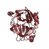
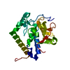
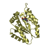
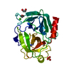
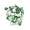
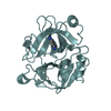
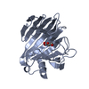
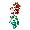
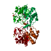
| タイトル | CRYSTAL STRUCTURE OF THE RNA TRIPHOSPHATASE DOMAIN OF MOUSE MRNA CAPPING ENZYME |
|---|
 要素 要素 | MRNA CAPPING ENZYME |
|---|
 キーワード キーワード | HYDROLASE / RNA triphosphatase domain / mRNA capping enzyme |
|---|
| 機能・相同性 |  機能・相同性情報 機能・相同性情報
RNA Pol II CTD phosphorylation and interaction with CE / mRNA Capping / RNA guanylyltransferase activity / inorganic triphosphate phosphatase activity / mRNA 5'-phosphatase / mRNA 5'-triphosphate monophosphatase activity / 7-methylguanosine mRNA capping / RNA processing / phosphoprotein phosphatase activity / mRNA guanylyltransferase ...RNA Pol II CTD phosphorylation and interaction with CE / mRNA Capping / RNA guanylyltransferase activity / inorganic triphosphate phosphatase activity / mRNA 5'-phosphatase / mRNA 5'-triphosphate monophosphatase activity / 7-methylguanosine mRNA capping / RNA processing / phosphoprotein phosphatase activity / mRNA guanylyltransferase / mRNA guanylyltransferase activity / GTP binding / ATP binding / nucleus類似検索 - 分子機能 mRNA capping enzyme, bifunctional / mRNA capping enzyme, adenylation domain / mRNA capping enzyme, C-terminal / mRNA capping enzyme, catalytic domain / mRNA capping enzyme, C-terminal domain / : / Dual specificity phosphatase, catalytic domain / Dual specificity phosphatase, catalytic domain / Dual specificity protein phosphatase domain profile. / Dual specificity protein phosphatase domain ...mRNA capping enzyme, bifunctional / mRNA capping enzyme, adenylation domain / mRNA capping enzyme, C-terminal / mRNA capping enzyme, catalytic domain / mRNA capping enzyme, C-terminal domain / : / Dual specificity phosphatase, catalytic domain / Dual specificity phosphatase, catalytic domain / Dual specificity protein phosphatase domain profile. / Dual specificity protein phosphatase domain / Protein tyrosine phosphatase superfamily / Protein-Tyrosine Phosphatase; Chain A / Tyrosine specific protein phosphatases active site. / Protein-tyrosine phosphatase, active site / Tyrosine specific protein phosphatases domain profile. / Tyrosine-specific protein phosphatases domain / Protein-tyrosine phosphatase-like / Nucleic acid-binding, OB-fold / Alpha-Beta Complex / Alpha Beta類似検索 - ドメイン・相同性 CACODYLATE ION / ISOPROPYL ALCOHOL / mRNA-capping enzyme類似検索 - 構成要素 |
|---|
| 生物種 |   Mus musculus (ハツカネズミ) Mus musculus (ハツカネズミ) |
|---|
| 手法 |  X線回折 / シンクロトロン / 多波長異常分散 / 解像度: 1.65 Å X線回折 / シンクロトロン / 多波長異常分散 / 解像度: 1.65 Å |
|---|
 データ登録者 データ登録者 | Changela, A. / Ho, C.K. / Martins, A. / Shuman, S. / Mondragon, A. |
|---|
 引用 引用 | ジャーナル: EMBO J. / 年: 2001
タイトル: Structure and mechanism of the RNA triphosphatase component of mammalian mRNA capping enzyme.
著者: Changela, A. / Ho, C.K. / Martins, A. / Shuman, S. / Mondragon, A. |
|---|
| 履歴 | | 登録 | 2001年3月20日 | 登録サイト: RCSB / 処理サイト: RCSB |
|---|
| 改定 1.0 | 2001年5月23日 | Provider: repository / タイプ: Initial release |
|---|
| 改定 1.1 | 2008年4月27日 | Group: Version format compliance |
|---|
| 改定 1.2 | 2011年7月13日 | Group: Derived calculations / Version format compliance |
|---|
| 改定 1.3 | 2024年2月7日 | Group: Data collection / Database references / Derived calculations
カテゴリ: chem_comp_atom / chem_comp_bond ...chem_comp_atom / chem_comp_bond / database_2 / pdbx_struct_conn_angle / struct_conn / struct_site
Item: _database_2.pdbx_DOI / _database_2.pdbx_database_accession ..._database_2.pdbx_DOI / _database_2.pdbx_database_accession / _pdbx_struct_conn_angle.ptnr1_auth_comp_id / _pdbx_struct_conn_angle.ptnr1_auth_seq_id / _pdbx_struct_conn_angle.ptnr1_label_asym_id / _pdbx_struct_conn_angle.ptnr1_label_atom_id / _pdbx_struct_conn_angle.ptnr1_label_comp_id / _pdbx_struct_conn_angle.ptnr1_label_seq_id / _pdbx_struct_conn_angle.ptnr1_symmetry / _pdbx_struct_conn_angle.ptnr2_auth_comp_id / _pdbx_struct_conn_angle.ptnr2_auth_seq_id / _pdbx_struct_conn_angle.ptnr2_label_asym_id / _pdbx_struct_conn_angle.ptnr2_label_atom_id / _pdbx_struct_conn_angle.ptnr2_label_comp_id / _pdbx_struct_conn_angle.ptnr3_auth_comp_id / _pdbx_struct_conn_angle.ptnr3_auth_seq_id / _pdbx_struct_conn_angle.ptnr3_label_asym_id / _pdbx_struct_conn_angle.ptnr3_label_atom_id / _pdbx_struct_conn_angle.ptnr3_label_comp_id / _pdbx_struct_conn_angle.ptnr3_symmetry / _pdbx_struct_conn_angle.value / _struct_conn.pdbx_dist_value / _struct_conn.ptnr1_auth_comp_id / _struct_conn.ptnr1_auth_seq_id / _struct_conn.ptnr1_label_asym_id / _struct_conn.ptnr1_label_atom_id / _struct_conn.ptnr1_label_comp_id / _struct_conn.ptnr1_label_seq_id / _struct_conn.ptnr2_auth_comp_id / _struct_conn.ptnr2_auth_seq_id / _struct_conn.ptnr2_label_asym_id / _struct_conn.ptnr2_label_atom_id / _struct_conn.ptnr2_label_comp_id / _struct_conn.ptnr2_label_seq_id / _struct_conn.ptnr2_symmetry / _struct_site.pdbx_auth_asym_id / _struct_site.pdbx_auth_comp_id / _struct_site.pdbx_auth_seq_id |
|---|
|
|---|
 ムービー
ムービー コントローラー


 データを開く
 基本情報
基本情報 構造の表示
構造の表示 ダウンロードとリンク
 その他のダウンロード

 PDBj
PDBj


 集合体
集合体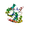


 分子量: 96.063 Da / 分子数: 1 / 由来タイプ: 合成 / 式: SO4
 分子量: 136.989 Da / 分子数: 1 / 由来タイプ: 合成 / 式: C2H6AsO2
 分子量: 24.305 Da / 分子数: 1 / 由来タイプ: 合成 / 式: Mg
 分子量: 60.095 Da / 分子数: 2 / 由来タイプ: 合成 / 式: C3H8O
 試料調製
試料調製 / ビームライン: BL9-1 / 波長: 0.97 Å
 解析
解析