 Movie
Movie Controller

[English] 日本語
 Yorodumi
- PDB-3r1b: Open crystal structure of cytochrome P450 2B4 covalently bound to... -
+ Open data

- Basic information
| Entry | Database: PDB / ID: 3r1b | |||||||||
|---|---|---|---|---|---|---|---|---|---|---|
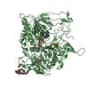
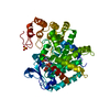
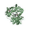
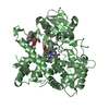
| Title | Open crystal structure of cytochrome P450 2B4 covalently bound to the mechanism-based inactivator tert-butylphenylacetylene | |||||||||
 Components Components | Cytochrome P450 2B4 | |||||||||
 Keywords Keywords | OXIDOREDUCTASE/OXIDOREDUCTASE INHIBITOR / cytochrome P450 2B4 / monooxygenase / membrane protein / OXIDOREDUCTASE-OXIDOREDUCTASE INHIBITOR complex | |||||||||
| Function / homology |  Function and homology information Function and homology informationarachidonate epoxygenase activity / epoxygenase P450 pathway / oxidoreductase activity, acting on paired donors, with incorporation or reduction of molecular oxygen, reduced flavin or flavoprotein as one donor, and incorporation of one atom of oxygen / unspecific monooxygenase / xenobiotic metabolic process / iron ion binding / heme binding / endoplasmic reticulum membrane Similarity search - Function | |||||||||
| Biological species |  | |||||||||
| Method |  X-RAY DIFFRACTION / SYNCHROTRON / MOLECULAR REPLACEMENT / molecular replacement / Resolution: 3 Å X-RAY DIFFRACTION / SYNCHROTRON / MOLECULAR REPLACEMENT / molecular replacement / Resolution: 3 Å | |||||||||
 Authors Authors | Gay, S.C. / Zhang, H. / Stout, C.D. / Hollenberg, P.F. / Halpert, J.R. | |||||||||
 Citation Citation | Journal: Biochemistry / Year: 2011 Title: Structural Analysis of Mammalian Cytochrome P450 2B4 Covalently Bound to the Mechanism-Based Inactivator tert-Butylphenylacetylene: Insight into Partial Enzymatic Activity. Authors: Gay, S.C. / Zhang, H. / Wilderman, P.R. / Roberts, A.G. / Liu, T. / Li, S. / Lin, H.L. / Zhang, Q. / Woods, V.L. / Stout, C.D. / Hollenberg, P.F. / Halpert, J.R. #1: Journal: J.Biol.Chem. / Year: 2006Title: Structure of microsomal cytochrome P450 2B4 complexed with the antifungal drug bifonazole: insight into P450 conformational plasticity and membrane interaction. Authors: Zhao, Y. / White, M.A. / Muralidhara, B.K. / Sun, L. / Halpert, J.R. / Stout, C.D. #2: Journal: J.Biol.Chem. / Year: 2004Title: Structure of mammalian cytochrome P450 2B4 complexed with 4-(4-chlorophenyl)imidazole at 1.9-A resolution: insight into the range of P450 conformations and the coordination of redox partner binding. Authors: Scott, E.E. / White, M.A. / He, Y.A. / Johnson, E.F. / Stout, C.D. / Halpert, J.R. #3: Journal: Proc.Natl.Acad.Sci.USA / Year: 2003Title: An open conformation of mammalian cytochrome P450 2B4 at 1.6-A resolution. Authors: Scott, E.E. / He, Y.A. / Wester, M.R. / White, M.A. / Chin, C.C. / Halpert, J.R. / Johnson, E.F. / Stout, C.D. #4: Journal: Biochemistry / Year: 2007Title: Structural and thermodynamic consequences of 1-(4-chlorophenyl)imidazole binding to cytochrome P450 2B4. Authors: Zhao, Y. / Sun, L. / Muralidhara, B.K. / Kumar, S. / White, M.A. / Stout, C.D. / Halpert, J.R. #5: Journal: Biochemistry / Year: 2009Title: Crystal structures of cytochrome P450 2B4 in complex with the inhibitor 1-biphenyl-4-methyl-1H-imidazole: ligand-induced structural response through alpha-helical repositioning. Authors: Gay, S.C. / Sun, L. / Maekawa, K. / Halpert, J.R. / Stout, C.D. #6: Journal: Biochemistry / Year: 2010Title: Structures of cytochrome P450 2B4 complexed with the antiplatelet drugs ticlopidine and clopidogrel . Authors: Gay, S.C. / Roberts, A.G. / Maekawa, K. / Talakad, J.C. / Hong, W.X. / Zhang, Q. / Stout, C.D. / Halpert, J.R. #7: Journal: J.Biol.Chem. / Year: 2010Title: Plasticity of cytochrome P450 2B4 as investigated by hydrogen-deuterium exchange mass spectrometry and X-ray crystallography. Authors: Wilderman, P.R. / Shah, M.B. / Liu, T. / Li, S. / Hsu, S. / Roberts, A.G. / Goodlett, D.R. / Zhang, Q. / Woods, V.L. / Stout, C.D. / Halpert, J.R. | |||||||||
| History |
|
- Structure visualization
| Structure viewer | Molecule: MolmilJmol/JSmol |
|---|
- Downloads & links
-Download
| PDBx/mmCIF format | 3r1b.cif.gz | 373.8 KB | Display | PDBx/mmCIF format |
|---|---|---|---|---|
| PDB format | pdb3r1b.ent.gz | 300.7 KB | Display | PDB format |
| PDBx/mmJSON format | 3r1b.json.gz | Tree view | PDBx/mmJSON format | |
| Others |  Other downloads Other downloads |
-Validation report
| Arichive directory | https://data.pdbj.org/pub/pdb/validation_reports/r1/3r1bftp://data.pdbj.org/pub/pdb/validation_reports/r1/3r1b | HTTPS FTP |
|---|
-Related structure data
| Related structure data |  3r1aC  1suoS C: citing same article ( S: Starting model for refinement |
|---|---|
| Similar structure data |


-Links
 PDBj
PDBj


- Assembly
| Deposited unit | 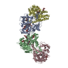
| ||||||||
|---|---|---|---|---|---|---|---|---|---|
| 1 | 
| ||||||||
| 2 | 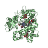
| ||||||||
| 3 | 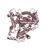
| ||||||||
| 4 | 
| ||||||||
| 5 | 
| ||||||||
| 6 | 
| ||||||||
| Unit cell |
|
-Components
| #1: Protein | Mass: 54169.082 Da / Num. of mol.: 4 Mutation: E2A, G22K, H23K, P24T, K25S, A26S, H27K, R29K, H226Y Source method: isolated from a genetically manipulated source Source: (gene. exp.)  #2: Polysaccharide | beta-D-fructofuranose-(2-1)-alpha-D-glucopyranose / sucrose   Source method: isolated from a genetically manipulated source Details: oligosaccharide with reducing-end-to-reducing-end glycosidic bond References: sucrose #3: Chemical | ChemComp-HEM /   Mass: 616.487 Da / Num. of mol.: 4 / Source method: obtained synthetically / Formula: C34H32FeN4O4 Mass: 616.487 Da / Num. of mol.: 4 / Source method: obtained synthetically / Formula: C34H32FeN4O4#4: Chemical | ChemComp-TB2 / (   Mass: 176.255 Da / Num. of mol.: 4 / Source method: obtained synthetically / Formula: C12H16O Mass: 176.255 Da / Num. of mol.: 4 / Source method: obtained synthetically / Formula: C12H16O#5: Chemical | ChemComp-CM5 /   Mass: 494.573 Da / Num. of mol.: 5 / Source method: obtained synthetically / Formula: C23H42O11 / Comment: detergent*YM Mass: 494.573 Da / Num. of mol.: 5 / Source method: obtained synthetically / Formula: C23H42O11 / Comment: detergent*YMHas protein modification | Y | Nonpolymer details | THE PROTEIN WAS INCUBATED WITH TERT-BUTYLPHENYLACETYLENE, CATALASE, CYTOCHROME P450 REDUCTASE, AND ...THE PROTEIN WAS INCUBATED WITH TERT-BUTYLPHENY | |
|---|
-Experimental details
-Experiment
| Experiment | Method: X-RAY DIFFRACTION / Number of used crystals: 1 |
|---|
- Sample preparation
| Crystal | Density Matthews: 3.77 Å3/Da / Density % sol: 67.39 % |
|---|---|
| Crystal grow | Temperature: 291 K / Method: vapor diffusion, sitting drop / pH: 7.5 Details: 0.1 M HEPES, 12% (w/v) PEG 3350, pH 7.5, vapor diffusion, sitting drop, temperature 291K |
-Data collection
| Diffraction | Mean temperature: 100 K | ||||||||||||||||||||||||||||||||||||||||||||||||||||||||||||||||||
|---|---|---|---|---|---|---|---|---|---|---|---|---|---|---|---|---|---|---|---|---|---|---|---|---|---|---|---|---|---|---|---|---|---|---|---|---|---|---|---|---|---|---|---|---|---|---|---|---|---|---|---|---|---|---|---|---|---|---|---|---|---|---|---|---|---|---|---|
| Diffraction source | Source: SYNCHROTRON / Site: SSRL  / Beamline: BL11-1 / Wavelength: 0.98 Å / Beamline: BL11-1 / Wavelength: 0.98 Å | ||||||||||||||||||||||||||||||||||||||||||||||||||||||||||||||||||
| Detector | Type: MARMOSAIC 325 mm CCD / Detector: CCD / Date: May 22, 2010 | ||||||||||||||||||||||||||||||||||||||||||||||||||||||||||||||||||
| Radiation | Monochromator: Si(111) / Protocol: SINGLE WAVELENGTH / Monochromatic (M) / Laue (L): M / Scattering type: x-ray | ||||||||||||||||||||||||||||||||||||||||||||||||||||||||||||||||||
| Radiation wavelength | Wavelength: 0.98 Å / Relative weight: 1 | ||||||||||||||||||||||||||||||||||||||||||||||||||||||||||||||||||
| Reflection | Resolution: 3→112.161 Å / Num. all: 63932 / Num. obs: 63932 / % possible obs: 99.4 % / Observed criterion σ(F): 0 / Observed criterion σ(I): 0 / Redundancy: 3.2 % / Biso Wilson estimate: 84.22 Å2 / Rmerge(I) obs: 0.092 / Rsym value: 0.092 / Net I/σ(I): 7.5 | ||||||||||||||||||||||||||||||||||||||||||||||||||||||||||||||||||
| Reflection shell |
|
-Phasing
| Phasing | Method: molecular replacement | |||||||||
|---|---|---|---|---|---|---|---|---|---|---|
| Phasing MR | Rfactor: 51.63 / Model details: Phaser MODE: MR_AUTO
|
- Processing
| Software |
| |||||||||||||||||||||||||||||||||||||||||||||||||||||||||||||||||||||||||||||||||||||||||||||||||||||||||||||||||||||||||||||||||||||||||||||||||||||||||||||||||
|---|---|---|---|---|---|---|---|---|---|---|---|---|---|---|---|---|---|---|---|---|---|---|---|---|---|---|---|---|---|---|---|---|---|---|---|---|---|---|---|---|---|---|---|---|---|---|---|---|---|---|---|---|---|---|---|---|---|---|---|---|---|---|---|---|---|---|---|---|---|---|---|---|---|---|---|---|---|---|---|---|---|---|---|---|---|---|---|---|---|---|---|---|---|---|---|---|---|---|---|---|---|---|---|---|---|---|---|---|---|---|---|---|---|---|---|---|---|---|---|---|---|---|---|---|---|---|---|---|---|---|---|---|---|---|---|---|---|---|---|---|---|---|---|---|---|---|---|---|---|---|---|---|---|---|---|---|---|---|---|---|---|---|
| Refinement | Method to determine structure: MOLECULAR REPLACEMENT Starting model: PDB ENTRY 1SUO Resolution: 3→53.43 Å / Occupancy max: 1 / Occupancy min: 0 / SU ML: 0.44 / σ(F): 0 / Stereochemistry target values: ML
| |||||||||||||||||||||||||||||||||||||||||||||||||||||||||||||||||||||||||||||||||||||||||||||||||||||||||||||||||||||||||||||||||||||||||||||||||||||||||||||||||
| Solvent computation | Shrinkage radii: 0.72 Å / VDW probe radii: 1 Å / Solvent model: FLAT BULK SOLVENT MODEL / Bsol: 81.04 Å2 / ksol: 0.33 e/Å3 | |||||||||||||||||||||||||||||||||||||||||||||||||||||||||||||||||||||||||||||||||||||||||||||||||||||||||||||||||||||||||||||||||||||||||||||||||||||||||||||||||
| Displacement parameters | Biso mean: 100.79 Å2
| |||||||||||||||||||||||||||||||||||||||||||||||||||||||||||||||||||||||||||||||||||||||||||||||||||||||||||||||||||||||||||||||||||||||||||||||||||||||||||||||||
| Refinement step | Cycle: LAST / Resolution: 3→53.43 Å
| |||||||||||||||||||||||||||||||||||||||||||||||||||||||||||||||||||||||||||||||||||||||||||||||||||||||||||||||||||||||||||||||||||||||||||||||||||||||||||||||||
| Refine LS restraints |
| |||||||||||||||||||||||||||||||||||||||||||||||||||||||||||||||||||||||||||||||||||||||||||||||||||||||||||||||||||||||||||||||||||||||||||||||||||||||||||||||||
| LS refinement shell |
|
