 Movie
Movie Controller

[English] 日本語
 Yorodumi
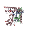
- PDB-2aj4: Crystal structure of Saccharomyces cerevisiae Galactokinase in co... -
+ Open data

- Basic information
| Entry | Database: PDB / ID: 2aj4 | ||||||
|---|---|---|---|---|---|---|---|
| Title | Crystal structure of Saccharomyces cerevisiae Galactokinase in complex with galactose and Mg:AMPPNP | ||||||
 Components Components | Galactokinase | ||||||
 Keywords Keywords | TRANSFERASE / galactokinase / galactosemia / transcription | ||||||
| Function / homology |  Function and homology information Function and homology informationpositive regulation of transcription by galactose / galactokinase / galactokinase activity / carbohydrate phosphorylation / beta-D-galactose catabolic process via UDP-galactose, Leloir pathway / galactose metabolic process / positive regulation of transcription from RNA polymerase II promoter by galactose / transcription regulator complex / ATP binding / nucleus ...positive regulation of transcription by galactose / galactokinase / galactokinase activity / carbohydrate phosphorylation / beta-D-galactose catabolic process via UDP-galactose, Leloir pathway / galactose metabolic process / positive regulation of transcription from RNA polymerase II promoter by galactose / transcription regulator complex / ATP binding / nucleus / cytoplasm / cytosol Similarity search - Function | ||||||
| Biological species |  | ||||||
| Method |  X-RAY DIFFRACTION / MOLECULAR REPLACEMENT / Resolution: 2.4 Å X-RAY DIFFRACTION / MOLECULAR REPLACEMENT / Resolution: 2.4 Å | ||||||
 Authors Authors | Thoden, J.B. / Sellick, C.A. / Timson, D.J. / Reece, R.J. / Holden, H.M. | ||||||
 Citation Citation | Journal: J.Biol.Chem. / Year: 2005 Title: Molecular structure of Saccharomyces cerevisiae Gal1p, a bifunctional galactokinase and transcriptional inducer Authors: Thoden, J.B. / Sellick, C.A. / Timson, D.J. / Reece, R.J. / Holden, H.M. | ||||||
| History |
|
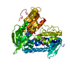
- Structure visualization
| Structure viewer | Molecule: MolmilJmol/JSmol |
|---|
- Downloads & links
-Download
| PDBx/mmCIF format | 2aj4.cif.gz | 212.8 KB | Display | PDBx/mmCIF format |
|---|---|---|---|---|
| PDB format | pdb2aj4.ent.gz | 167.4 KB | Display | PDB format |
| PDBx/mmJSON format | 2aj4.json.gz | Tree view | PDBx/mmJSON format | |
| Others |  Other downloads Other downloads |
-Validation report
| Arichive directory | https://data.pdbj.org/pub/pdb/validation_reports/aj/2aj4ftp://data.pdbj.org/pub/pdb/validation_reports/aj/2aj4 | HTTPS FTP |
|---|
-Related structure data
| Related structure data |  2a2cS S: Starting model for refinement |
|---|---|
| Similar structure data |


-Links
 PDBj
PDBj


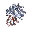
- Assembly
| Deposited unit | 
| ||||||||
|---|---|---|---|---|---|---|---|---|---|
| 1 | 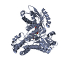
| ||||||||
| 2 | 
| ||||||||
| Unit cell |
|
-Components
-Protein / Sugars , 2 types, 4 molecules AB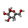
| #1: Protein | Mass: 60364.594 Da / Num. of mol.: 2 Source method: isolated from a genetically manipulated source Source: (gene. exp.) Gene: GAL1 / Plasmid: pET28 / Production host:  #2: Sugar |  Type: D-saccharide, alpha linking / Mass: 180.156 Da / Num. of mol.: 2 Type: D-saccharide, alpha linking / Mass: 180.156 Da / Num. of mol.: 2Source method: isolated from a genetically manipulated source Formula: C6H12O6 |
|---|
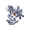
-Non-polymers , 4 types, 197 molecules 


| #3: Chemical |  Mass: 24.305 Da / Num. of mol.: 2 / Source method: obtained synthetically / Formula: Mg Mass: 24.305 Da / Num. of mol.: 2 / Source method: obtained synthetically / Formula: Mg#4: Chemical |  Mass: 35.453 Da / Num. of mol.: 2 / Source method: obtained synthetically / Formula: Cl Mass: 35.453 Da / Num. of mol.: 2 / Source method: obtained synthetically / Formula: Cl#5: Chemical |  Mass: 506.196 Da / Num. of mol.: 2 / Source method: obtained synthetically / Formula: C10H17N6O12P3 / Comment: AMP-PNP, energy-carrying molecule analogue*YM Mass: 506.196 Da / Num. of mol.: 2 / Source method: obtained synthetically / Formula: C10H17N6O12P3 / Comment: AMP-PNP, energy-carrying molecule analogue*YM#6: Water | ChemComp-HOH / | Mass: 18.015 Da / Num. of mol.: 191 / Source method: isolated from a natural source / Formula: H2O |
|---|
-Experimental details
-Experiment
| Experiment | Method: X-RAY DIFFRACTION / Number of used crystals: 1 |
|---|
- Sample preparation
| Crystal | Density Matthews: 2.5 Å3/Da / Density % sol: 50 % |
|---|---|
| Crystal grow | Temperature: 294 K / Method: vapor diffusion, hanging drop / pH: 6 Details: PEG8000, LiCl, galactose, Mg:AMPPNP, MES, pH 6.0, VAPOR DIFFUSION, HANGING DROP, temperature 294K |
-Data collection
| Diffraction | Mean temperature: 290 K |
|---|---|
| Diffraction source | Source: ROTATING ANODE / Type: RIGAKU RU200 / Wavelength: 1.5418 Å |
| Detector | Type: Bruker Platinum 135 / Detector: CCD / Date: Mar 6, 2005 / Details: montel |
| Radiation | Monochromator: Ni FILTER / Protocol: SINGLE WAVELENGTH / Monochromatic (M) / Laue (L): M / Scattering type: x-ray |
| Radiation wavelength | Wavelength: 1.5418 Å / Relative weight: 1 |
| Reflection | Resolution: 2.4→30 Å / Num. all: 44350 / Num. obs: 44350 / % possible obs: 95.5 % / Observed criterion σ(F): 0 / Observed criterion σ(I): 0 / Redundancy: 3.4 % / Rsym value: 0.075 / Net I/σ(I): 11.2 |
| Reflection shell | Resolution: 2.4→2.5 Å / Redundancy: 1.7 % / Mean I/σ(I) obs: 2.9 / Num. unique all: 4591 / Rsym value: 0.273 / % possible all: 86 |
- Processing
| Software |
| |||||||||||||||||||||||||
|---|---|---|---|---|---|---|---|---|---|---|---|---|---|---|---|---|---|---|---|---|---|---|---|---|---|---|
| Refinement | Method to determine structure: MOLECULAR REPLACEMENT Starting model: PDB entry 2A2C Resolution: 2.4→30 Å / Cross valid method: THROUGHOUT / σ(F): 0 / Stereochemistry target values: Engh & Huber
| |||||||||||||||||||||||||
| Refinement step | Cycle: LAST / Resolution: 2.4→30 Å
| |||||||||||||||||||||||||
| Refine LS restraints |
|
