 Movie
Movie Controller

[English] 日本語
 Yorodumi
- PDB-6o7c: Crystal structure of the LjCASTOR gating ring in the Ca2+ and K+ state -
+ Open data

- Basic information
| Entry | Database: PDB / ID: 6o7c | ||||||||||||
|---|---|---|---|---|---|---|---|---|---|---|---|---|---|
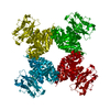
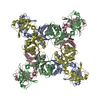
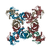
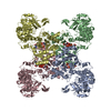
| Title | Crystal structure of the LjCASTOR gating ring in the Ca2+ and K+ state | ||||||||||||
 Components Components | Ion channel CASTOR | ||||||||||||
 Keywords Keywords | TRANSPORT PROTEIN / calcium channel / RCK domain / gating ring / CASTOR / membrane protein | ||||||||||||
| Function / homology |  Function and homology information Function and homology informationpotassium ion transport / nuclear membrane / monoatomic ion transmembrane transport Similarity search - Function | ||||||||||||
| Biological species |  | ||||||||||||
| Method |  X-RAY DIFFRACTION / SYNCHROTRON / SAD / Resolution: 1.85 Å X-RAY DIFFRACTION / SYNCHROTRON / SAD / Resolution: 1.85 Å | ||||||||||||
 Authors Authors | Jiang, Y. / Kim, S. | ||||||||||||
| Funding support |  United States, 3items United States, 3items
| ||||||||||||
 Citation Citation | Journal: Nat Commun / Year: 2019 Title: Ca2+-regulated Ca2+channels with an RCK gating ring control plant symbiotic associations. Authors: Kim, S. / Zeng, W. / Bernard, S. / Liao, J. / Venkateshwaran, M. / Ane, J.M. / Jiang, Y. | ||||||||||||
| History |
|
- Structure visualization
| Structure viewer | Molecule: MolmilJmol/JSmol |
|---|
- Downloads & links
-Download
| PDBx/mmCIF format | 6o7c.cif.gz | 263.6 KB | Display | PDBx/mmCIF format |
|---|---|---|---|---|
| PDB format | pdb6o7c.ent.gz | 176 KB | Display | PDB format |
| PDBx/mmJSON format | 6o7c.json.gz | Tree view | PDBx/mmJSON format | |
| Others |  Other downloads Other downloads |
-Validation report
| Arichive directory | https://data.pdbj.org/pub/pdb/validation_reports/o7/6o7cftp://data.pdbj.org/pub/pdb/validation_reports/o7/6o7c | HTTPS FTP |
|---|
-Related structure data


-Links
 PDBj
PDBj
- Assembly
| Deposited unit | 
| ||||||||||
|---|---|---|---|---|---|---|---|---|---|---|---|
| 1 | 
| ||||||||||
| Unit cell |
|
-Components
-Protein , 1 types, 1 molecules A
| #1: Protein | Mass: 60537.285 Da / Num. of mol.: 1 / Fragment: gating ring (UNP residues 312-853) Source method: isolated from a genetically manipulated source Source: (gene. exp.)  |
|---|
-Non-polymers , 5 types, 299 molecules 


| #2: Chemical |  Mass: 40.078 Da / Num. of mol.: 3 / Source method: obtained synthetically / Formula: Ca / Feature type: SUBJECT OF INVESTIGATION Mass: 40.078 Da / Num. of mol.: 3 / Source method: obtained synthetically / Formula: Ca / Feature type: SUBJECT OF INVESTIGATION#3: Chemical | ChemComp-MG / |  Mass: 24.305 Da / Num. of mol.: 1 / Source method: obtained synthetically / Formula: Mg / Feature type: SUBJECT OF INVESTIGATION Mass: 24.305 Da / Num. of mol.: 1 / Source method: obtained synthetically / Formula: Mg / Feature type: SUBJECT OF INVESTIGATION#4: Chemical | ChemComp-K / |  Mass: 39.098 Da / Num. of mol.: 1 / Source method: obtained synthetically / Formula: K / Feature type: SUBJECT OF INVESTIGATION Mass: 39.098 Da / Num. of mol.: 1 / Source method: obtained synthetically / Formula: K / Feature type: SUBJECT OF INVESTIGATION#5: Chemical | ChemComp-NA / |  Mass: 22.990 Da / Num. of mol.: 1 / Source method: obtained synthetically / Formula: Na / Feature type: SUBJECT OF INVESTIGATION Mass: 22.990 Da / Num. of mol.: 1 / Source method: obtained synthetically / Formula: Na / Feature type: SUBJECT OF INVESTIGATION#6: Water | ChemComp-HOH / | Mass: 18.015 Da / Num. of mol.: 293 / Source method: isolated from a natural source / Formula: H2O |
|---|
-Experimental details
-Experiment
| Experiment | Method: X-RAY DIFFRACTION / Number of used crystals: 1 |
|---|
- Sample preparation
| Crystal | Density Matthews: 2.7 Å3/Da / Density % sol: 54.37 % / Description: Thin rectangular prism with smooth edge |
|---|---|
| Crystal grow | Temperature: 293 K / Method: vapor diffusion, sitting drop / pH: 8.2 Details: 21% w/v PEG550 MME, 50 mM magnesium acetate, 100 mM Tris, pH 8.2 |
-Data collection
| Diffraction | Mean temperature: 100 K / Serial crystal experiment: N |
|---|---|
| Diffraction source | Source: SYNCHROTRON / Site: ALS / Beamline: 8.2.2 / Wavelength: 0.99996 Å |
| Detector | Type: ADSC QUANTUM 315r / Detector: CCD / Date: May 20, 2016 |
| Radiation | Monochromator: double crystal Si(111) / Protocol: SINGLE WAVELENGTH / Monochromatic (M) / Laue (L): M / Scattering type: x-ray |
| Radiation wavelength | Wavelength: 0.99996 Å / Relative weight: 1 |
| Reflection | Resolution: 1.85→50 Å / Num. obs: 55045 / % possible obs: 99.7 % / Redundancy: 7.4 % / Biso Wilson estimate: 39.01 Å2 / Rmerge(I) obs: 0.063 / Rpim(I) all: 0.025 / Rrim(I) all: 0.068 / Rsym value: 0.063 / Net I/av σ(I): 42.512 / Net I/σ(I): 23.4 |
| Reflection shell | Resolution: 1.85→1.88 Å / Redundancy: 7.2 % / Rmerge(I) obs: 1.67 / Mean I/σ(I) obs: 1 / Num. unique obs: 2759 / CC1/2: 0.69 / Rpim(I) all: 0.668 / Rrim(I) all: 1.799 / Rsym value: 1.67 / Χ2: 0.47 |
- Processing
| Software |
| |||||||||||||||||||||||||||||||||||||||||||||||||||||||||||||||||||||||||||||||||||||||||||||||||||||||||
|---|---|---|---|---|---|---|---|---|---|---|---|---|---|---|---|---|---|---|---|---|---|---|---|---|---|---|---|---|---|---|---|---|---|---|---|---|---|---|---|---|---|---|---|---|---|---|---|---|---|---|---|---|---|---|---|---|---|---|---|---|---|---|---|---|---|---|---|---|---|---|---|---|---|---|---|---|---|---|---|---|---|---|---|---|---|---|---|---|---|---|---|---|---|---|---|---|---|---|---|---|---|---|---|---|---|---|
| Refinement | Method to determine structure: SAD / Resolution: 1.85→39.71 Å / SU ML: 0.1896 / Cross valid method: FREE R-VALUE / σ(F): 1.34 / Phase error: 22.3623
| |||||||||||||||||||||||||||||||||||||||||||||||||||||||||||||||||||||||||||||||||||||||||||||||||||||||||
| Solvent computation | Shrinkage radii: 0.9 Å / VDW probe radii: 1.11 Å | |||||||||||||||||||||||||||||||||||||||||||||||||||||||||||||||||||||||||||||||||||||||||||||||||||||||||
| Displacement parameters | Biso mean: 49.12 Å2 | |||||||||||||||||||||||||||||||||||||||||||||||||||||||||||||||||||||||||||||||||||||||||||||||||||||||||
| Refinement step | Cycle: LAST / Resolution: 1.85→39.71 Å
| |||||||||||||||||||||||||||||||||||||||||||||||||||||||||||||||||||||||||||||||||||||||||||||||||||||||||
| Refine LS restraints |
| |||||||||||||||||||||||||||||||||||||||||||||||||||||||||||||||||||||||||||||||||||||||||||||||||||||||||
| LS refinement shell |
| |||||||||||||||||||||||||||||||||||||||||||||||||||||||||||||||||||||||||||||||||||||||||||||||||||||||||
| Refinement TLS params. | Method: refined / Refine-ID: X-RAY DIFFRACTION
| |||||||||||||||||||||||||||||||||||||||||||||||||||||||||||||||||||||||||||||||||||||||||||||||||||||||||
| Refinement TLS group |
|
