 Movie
Movie Controller

[English] 日本語
 Yorodumi
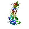
- PDB-3pry: Crystal structure of the middle domain of human HSP90-beta refine... -
+ Open data

- Basic information
| Entry | Database: PDB / ID: 3pry | ||||||
|---|---|---|---|---|---|---|---|
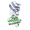
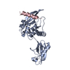
| Title | Crystal structure of the middle domain of human HSP90-beta refined at 2.3 A resolution | ||||||
 Components Components | Heat shock protein HSP 90-beta | ||||||
 Keywords Keywords | CHAPERONE / Structural Genomics / Structural Genomics Consortium / SGC / heat shock protein / HSP90B | ||||||
| Function / homology |  Function and homology information Function and homology informationHSP90-CDC37 chaperone complex / negative regulation of proteasomal protein catabolic process / Aryl hydrocarbon receptor signalling / aryl hydrocarbon receptor complex / histone methyltransferase binding / dynein axonemal particle / receptor ligand inhibitor activity / protein kinase regulator activity / positive regulation of protein localization to cell surface / ATP-dependent protein binding ...HSP90-CDC37 chaperone complex / negative regulation of proteasomal protein catabolic process / Aryl hydrocarbon receptor signalling / aryl hydrocarbon receptor complex / histone methyltransferase binding / dynein axonemal particle / receptor ligand inhibitor activity / protein kinase regulator activity / positive regulation of protein localization to cell surface / ATP-dependent protein binding / Respiratory syncytial virus genome replication / telomerase holoenzyme complex assembly / Uptake and function of diphtheria toxin / positive regulation of transforming growth factor beta receptor signaling pathway / dendritic growth cone / TPR domain binding / Assembly and release of respiratory syncytial virus (RSV) virions / Sema3A PAK dependent Axon repulsion / The NLRP3 inflammasome / protein phosphatase activator activity / regulation of protein ubiquitination / HSF1-dependent transactivation / protein folding chaperone complex / response to unfolded protein / Attenuation phase / HSF1 activation / chaperone-mediated protein complex assembly / RHOBTB2 GTPase cycle / axonal growth cone / telomere maintenance via telomerase / Purinergic signaling in leishmaniasis infection / supramolecular fiber organization / heat shock protein binding / peptide binding / DNA polymerase binding / protein folding chaperone / ESR-mediated signaling / negative regulation of proteasomal ubiquitin-dependent protein catabolic process / HSP90 chaperone cycle for steroid hormone receptors (SHR) in the presence of ligand / cellular response to interleukin-4 / placenta development / nitric-oxide synthase regulator activity / positive regulation of cell differentiation / ATP-dependent protein folding chaperone / Regulation of actin dynamics for phagocytic cup formation / DDX58/IFIH1-mediated induction of interferon-alpha/beta / tau protein binding / histone deacetylase binding / kinase binding / Chaperone Mediated Autophagy / disordered domain specific binding / positive regulation of nitric oxide biosynthetic process / : / The role of GTSE1 in G2/M progression after G2 checkpoint / melanosome / MHC class II protein complex binding / regulation of protein localization / double-stranded RNA binding / cellular response to heat / protein folding / secretory granule lumen / Estrogen-dependent gene expression / Potential therapeutics for SARS / ficolin-1-rich granule lumen / protein dimerization activity / regulation of cell cycle / protein stabilization / cadherin binding / neuronal cell body / Neutrophil degranulation / ubiquitin protein ligase binding / protein kinase binding / negative regulation of apoptotic process / virion attachment to host cell / perinuclear region of cytoplasm / SARS-CoV-2 activates/modulates innate and adaptive immune responses / cell surface / protein homodimerization activity / ATP hydrolysis activity / protein-containing complex / mitochondrion / RNA binding / extracellular exosome / extracellular region / nucleoplasm / ATP binding / membrane / identical protein binding / nucleus / plasma membrane / cytoplasm / cytosol Similarity search - Function | ||||||
| Biological species |  Homo sapiens (human) Homo sapiens (human) | ||||||
| Method |  X-RAY DIFFRACTION / SYNCHROTRON / MOLECULAR REPLACEMENT / Resolution: 2.28 Å X-RAY DIFFRACTION / SYNCHROTRON / MOLECULAR REPLACEMENT / Resolution: 2.28 Å | ||||||
 Authors Authors | Chaikuad, A. / Pilka, E. / Sharpe, T.D. / Cooper, C.D.O. / Phillips, C. / Berridge, G. / Ayinampudi, V. / Fedorov, O. / Keates, T. / Thangaratnarajah, C. ...Chaikuad, A. / Pilka, E. / Sharpe, T.D. / Cooper, C.D.O. / Phillips, C. / Berridge, G. / Ayinampudi, V. / Fedorov, O. / Keates, T. / Thangaratnarajah, C. / Zimmermann, T. / Vollmar, M. / Yue, W.W. / Che, K.H. / Krojer, T. / Muniz, J.R.C. / von Delft, F. / Bountra, C. / Arrowsmith, C.H. / Weigelt, J. / Edwards, A. / Bullock, A. / Structural Genomics Consortium (SGC) | ||||||
 Citation Citation | Journal: To be Published Title: Crystal structure of the middle domain of human HSP90-beta refined at 2.3 A resolution Authors: Chaikuad, A. / Pilka, E. / Sharpe, T.D. / Cooper, C.D.O. / Phillips, C. / Berridge, G. / Ayinampudi, V. / Fedorov, O. / Keates, T. / Thangaratnarajah, C. / Zimmermann, T. / Vollmar, M. / ...Authors: Chaikuad, A. / Pilka, E. / Sharpe, T.D. / Cooper, C.D.O. / Phillips, C. / Berridge, G. / Ayinampudi, V. / Fedorov, O. / Keates, T. / Thangaratnarajah, C. / Zimmermann, T. / Vollmar, M. / Yue, W.W. / Che, K.H. / Krojer, T. / Muniz, J.R.C. / von Delft, F. / Bountra, C. / Arrowsmith, C.H. / Weigelt, J. / Edwards, A. / Bullock, A. / Structural Genomics Consortium (SGC) | ||||||
| History |
|
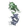
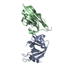
- Structure visualization
| Structure viewer | Molecule: MolmilJmol/JSmol |
|---|
- Downloads & links
-Download
| PDBx/mmCIF format | 3pry.cif.gz | 319.5 KB | Display | PDBx/mmCIF format |
|---|---|---|---|---|
| PDB format | pdb3pry.ent.gz | 260.9 KB | Display | PDB format |
| PDBx/mmJSON format | 3pry.json.gz | Tree view | PDBx/mmJSON format | |
| Others |  Other downloads Other downloads |
-Validation report
| Arichive directory | https://data.pdbj.org/pub/pdb/validation_reports/pr/3pryftp://data.pdbj.org/pub/pdb/validation_reports/pr/3pry | HTTPS FTP |
|---|
-Related structure data
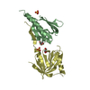
| Related structure data | 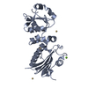 1hk7S S: Starting model for refinement |
|---|---|
| Similar structure data |


-Links
 PDBj
PDBj


- Assembly
| Deposited unit | 
| |||||||||||||||||||||||||||||||||||||||||||||||||||||||||||||||||||||||||||||||||||||||||||||||||||||||||||||||||||||||||||||||||||||||||||||||||||||||||||||||||||||||||||||||||||||||||||||||||||||||||||||
|---|---|---|---|---|---|---|---|---|---|---|---|---|---|---|---|---|---|---|---|---|---|---|---|---|---|---|---|---|---|---|---|---|---|---|---|---|---|---|---|---|---|---|---|---|---|---|---|---|---|---|---|---|---|---|---|---|---|---|---|---|---|---|---|---|---|---|---|---|---|---|---|---|---|---|---|---|---|---|---|---|---|---|---|---|---|---|---|---|---|---|---|---|---|---|---|---|---|---|---|---|---|---|---|---|---|---|---|---|---|---|---|---|---|---|---|---|---|---|---|---|---|---|---|---|---|---|---|---|---|---|---|---|---|---|---|---|---|---|---|---|---|---|---|---|---|---|---|---|---|---|---|---|---|---|---|---|---|---|---|---|---|---|---|---|---|---|---|---|---|---|---|---|---|---|---|---|---|---|---|---|---|---|---|---|---|---|---|---|---|---|---|---|---|---|---|---|---|---|---|---|---|---|---|---|---|---|
| 1 | 
| |||||||||||||||||||||||||||||||||||||||||||||||||||||||||||||||||||||||||||||||||||||||||||||||||||||||||||||||||||||||||||||||||||||||||||||||||||||||||||||||||||||||||||||||||||||||||||||||||||||||||||||
| 2 | 
| |||||||||||||||||||||||||||||||||||||||||||||||||||||||||||||||||||||||||||||||||||||||||||||||||||||||||||||||||||||||||||||||||||||||||||||||||||||||||||||||||||||||||||||||||||||||||||||||||||||||||||||
| 3 | 
| |||||||||||||||||||||||||||||||||||||||||||||||||||||||||||||||||||||||||||||||||||||||||||||||||||||||||||||||||||||||||||||||||||||||||||||||||||||||||||||||||||||||||||||||||||||||||||||||||||||||||||||
| Unit cell |
| |||||||||||||||||||||||||||||||||||||||||||||||||||||||||||||||||||||||||||||||||||||||||||||||||||||||||||||||||||||||||||||||||||||||||||||||||||||||||||||||||||||||||||||||||||||||||||||||||||||||||||||
| Noncrystallographic symmetry (NCS) | NCS domain:
NCS domain segments:
|