 Movie
Movie Controller

[English] 日本語
 Yorodumi
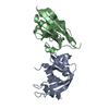
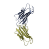
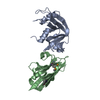
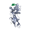
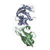
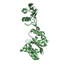
- PDB-5yca: Crystal structure of inner membrane protein Bqt4 in complex with LEM2 -
+ Open data

- Basic information
| Entry | Database: PDB / ID: 5yca | ||||||
|---|---|---|---|---|---|---|---|
| Title | Crystal structure of inner membrane protein Bqt4 in complex with LEM2 | ||||||
 Components Components |
| ||||||
 Keywords Keywords | MEMBRANE PROTEIN / Chromosome organization / Membrane organization / Bouquet formation / Lap-Emerin-Man domain protein | ||||||
| Function / homology |  Function and homology information Function and homology informationheterochromatin-nuclear membrane anchor activity / centromere-nuclear envelope anchor activity / subnuclear spatial organization of silent mating-type cassette heterochromatin / mitotic nuclear bridge midzone membrane domain / Sealing of the nuclear envelope (NE) by ESCRT-III / meiotic telomere tethering at nuclear periphery / nuclear membrane biogenesis / telomere-nuclear envelope anchor activity / nuclear membrane complex Bqt3-Bqt4 / centromere clustering at the mitotic interphase nuclear envelope ...heterochromatin-nuclear membrane anchor activity / centromere-nuclear envelope anchor activity / subnuclear spatial organization of silent mating-type cassette heterochromatin / mitotic nuclear bridge midzone membrane domain / Sealing of the nuclear envelope (NE) by ESCRT-III / meiotic telomere tethering at nuclear periphery / nuclear membrane biogenesis / telomere-nuclear envelope anchor activity / nuclear membrane complex Bqt3-Bqt4 / centromere clustering at the mitotic interphase nuclear envelope / attachment of telomeric heterochromatin to nuclear envelope / CENP-A containing chromatin / regulation of membrane lipid distribution / mitotic telomere tethering at nuclear periphery / : / protein localization to heterochromatin / chromosome, centromeric core domain / nuclear membrane organization / meiotic attachment of telomere to nuclear envelope / nuclear inner membrane organization / chromosome, subtelomeric region / mitotic spindle pole body / meiotic telomere clustering / heterochromatin organization / SUMO is conjugated to E1 (UBA2:SAE1) / SUMOylation of nuclear envelope proteins / SUMO is transferred from E1 to E2 (UBE2I, UBC9) / SUMO is proteolytically processed / nuclear envelope organization / SUMOylation of transcription factors / centromeric DNA binding / Postmitotic nuclear pore complex (NPC) reformation / SUMOylation of transcription cofactors / septin ring / mitotic nuclear membrane reassembly / SUMOylation of DNA damage response and repair proteins / Transcriptional and post-translational regulation of MITF-M expression and activity / SUMOylation of DNA replication proteins / rDNA heterochromatin / phosphatidic acid binding / SUMOylation of SUMOylation proteins / Recruitment and ATM-mediated phosphorylation of repair and signaling proteins at DNA double strand breaks / SUMOylation of RNA binding proteins / nuclear inner membrane / SUMOylation of chromatin organization proteins / ubiquitin-like protein ligase binding / protein sumoylation / chromosome organization / pericentric heterochromatin / nuclear periphery / telomere organization / telomere maintenance / condensed nuclear chromosome / meiotic cell cycle / molecular condensate scaffold activity / protein tag activity / nuclear envelope / double-stranded DNA binding / nuclear membrane / cell division / chromatin binding / lipid binding / DNA binding / membrane / identical protein binding / nucleus / cytoplasm Similarity search - Function | ||||||
| Biological species |  | ||||||
| Method |  X-RAY DIFFRACTION / SYNCHROTRON / MOLECULAR REPLACEMENT / Resolution: 1.57 Å X-RAY DIFFRACTION / SYNCHROTRON / MOLECULAR REPLACEMENT / Resolution: 1.57 Å | ||||||
 Authors Authors | Chen, Y. / Hu, C. | ||||||
 Citation Citation | Journal: Nucleic Acids Res. / Year: 2019 Title: Structural insights into chromosome attachment to the nuclear envelope by an inner nuclear membrane protein Bqt4 in fission yeast. Authors: Hu, C. / Inoue, H. / Sun, W. / Takeshita, Y. / Huang, Y. / Xu, Y. / Kanoh, J. / Chen, Y. | ||||||
| History |
|
- Structure visualization
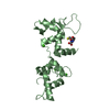
| Structure viewer | Molecule: MolmilJmol/JSmol |
|---|
- Downloads & links
-Download
| PDBx/mmCIF format | 5yca.cif.gz | 67.2 KB | Display | PDBx/mmCIF format |
|---|---|---|---|---|
| PDB format | pdb5yca.ent.gz | 47.3 KB | Display | PDB format |
| PDBx/mmJSON format | 5yca.json.gz | Tree view | PDBx/mmJSON format | |
| Others |  Other downloads Other downloads |
-Validation report
| Arichive directory | https://data.pdbj.org/pub/pdb/validation_reports/yc/5ycaftp://data.pdbj.org/pub/pdb/validation_reports/yc/5yca | HTTPS FTP |
|---|
-Related structure data
| Related structure data |  5yc2SC 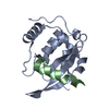 6a6wC 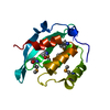 5ybxS S: Starting model for refinement C: citing same article ( |
|---|---|
| Similar structure data |


-Links
 PDBj
PDBj


- Assembly
| Deposited unit | 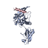
| ||||||||
|---|---|---|---|---|---|---|---|---|---|
| 1 |
| ||||||||
| Unit cell |
|
-Components
| #1: Protein | Mass: 23570.859 Da / Num. of mol.: 1 / Mutation: Q61E Source method: isolated from a genetically manipulated source Details: SUMO (residue 20 to 92) tagged Bqt4 (residue 8 to 140) Source: (gene. exp.) Strain: ATCC 204508 / S288c, 972 / ATCC 24843 / Gene: SMT3, YDR510W, D9719.15, bqt4, SPBC19C7.10 / Plasmid: pET28b-SUMO / Production host:  |
|---|---|
| #2: Protein/peptide | Mass: 2372.500 Da / Num. of mol.: 1 Source method: isolated from a genetically manipulated source Details: Bqt4 binding motif of LEM2 protein Source: (gene. exp.) Strain: 972 / ATCC 24843 / Gene: lem2, heh1, SPAC18G6.10 / Plasmid: pGEX-6P-1 / Production host: |
| #3: Water | ChemComp-HOH /  Mass: 18.015 Da / Num. of mol.: 294 / Source method: isolated from a natural source / Formula: H2O Mass: 18.015 Da / Num. of mol.: 294 / Source method: isolated from a natural source / Formula: H2O |
-Experimental details
-Experiment
| Experiment | Method: X-RAY DIFFRACTION / Number of used crystals: 1 |
|---|
- Sample preparation
| Crystal | Density Matthews: 2.51 Å3/Da / Density % sol: 50.96 % / Description: beautiful diamond |
|---|---|
| Crystal grow | Temperature: 289 K / Method: vapor diffusion, sitting drop / pH: 5.6 / Details: 1600 mM Sodium citrate tribasic |
-Data collection
| Diffraction | Mean temperature: 100 K / Ambient temp details: liquid nitrogen |
|---|---|
| Diffraction source | Source: SYNCHROTRON / Site: NFPSS  / Beamline: BL19U1 / Wavelength: 0.97853 Å / Beamline: BL19U1 / Wavelength: 0.97853 Å |
| Detector | Type: DECTRIS PILATUS3 6M / Detector: PIXEL / Date: Jun 10, 2017 |
| Radiation | Protocol: SINGLE WAVELENGTH / Monochromatic (M) / Laue (L): M / Scattering type: x-ray |
| Radiation wavelength | Wavelength: 0.97853 Å / Relative weight: 1 |
| Reflection | Resolution: 1.57→27.392 Å / Num. obs: 36534 / % possible obs: 99.9 % / Redundancy: 3 % / Biso Wilson estimate: 17.71 Å2 / Net I/σ(I): 3.7 |
| Reflection shell | Resolution: 1.57→1.6 Å / Redundancy: 11.6 % / Rmerge(I) obs: 0.264 / CC1/2: 0.978 / Rpim(I) all: 0.079 / Rrim(I) all: 0.276 / Χ2: 0.712 / % possible all: 98.6 |
- Processing
| Software |
| ||||||||||||||||||||||||||||||||||||||||||||||||||||||||||||||||||||||||||||||||||||
|---|---|---|---|---|---|---|---|---|---|---|---|---|---|---|---|---|---|---|---|---|---|---|---|---|---|---|---|---|---|---|---|---|---|---|---|---|---|---|---|---|---|---|---|---|---|---|---|---|---|---|---|---|---|---|---|---|---|---|---|---|---|---|---|---|---|---|---|---|---|---|---|---|---|---|---|---|---|---|---|---|---|---|---|---|---|
| Refinement | Method to determine structure: MOLECULAR REPLACEMENT Starting model: 5YBX, 5YC2 Resolution: 1.57→27.392 Å / SU ML: 0.14 / Cross valid method: FREE R-VALUE / σ(F): 1.35 / Phase error: 20.69
| ||||||||||||||||||||||||||||||||||||||||||||||||||||||||||||||||||||||||||||||||||||
| Solvent computation | Shrinkage radii: 0.9 Å / VDW probe radii: 1.11 Å | ||||||||||||||||||||||||||||||||||||||||||||||||||||||||||||||||||||||||||||||||||||
| Displacement parameters | Biso max: 66.96 Å2 / Biso mean: 23.33 Å2 / Biso min: 10.14 Å2 | ||||||||||||||||||||||||||||||||||||||||||||||||||||||||||||||||||||||||||||||||||||
| Refinement step | Cycle: final / Resolution: 1.57→27.392 Å
| ||||||||||||||||||||||||||||||||||||||||||||||||||||||||||||||||||||||||||||||||||||
| Refine LS restraints |
| ||||||||||||||||||||||||||||||||||||||||||||||||||||||||||||||||||||||||||||||||||||
| LS refinement shell | Refine-ID: X-RAY DIFFRACTION / Rfactor Rfree error: 0
|
