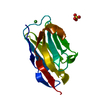
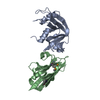
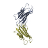
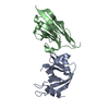
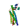
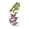
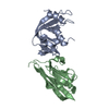
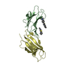
Mass: 13708.326 Da / Num. of mol.: 1 / Source method: isolated from a natural source / Source: (natural) Bos taurus (domestic cattle) / References: UniProt: P61823, EC: 3.1.27.5
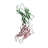
#2: Antibody
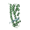
VHHAntibody
Mass: 12998.415 Da / Num. of mol.: 1 Source method: isolated from a genetically manipulated source Source: (gene. exp.) Lama glama (llama) / Production host: Escherichia coli (E. coli)
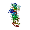
Method to determine structure: MOLECULAR REPLACEMENT / Resolution: 1.75→21.29 Å / Cor.coef. Fo:Fc: 0.951 / Cor.coef. Fo:Fc free: 0.927 / SU B: 2.794 / SU ML: 0.091 / Cross valid method: THROUGHOUT / σ(F): 2 / ESU R: 0.15 / ESU R Free: 0.139 / Stereochemistry target values: MAXIMUM LIKELIHOOD / Details: HYDROGENS HAVE BEEN USED IF PRESENT IN THE INPUT
Rfactor
Num. reflection
% reflection
Selection details
Rfree
0.22987
890
5 %
RANDOM
Rwork
0.18214
-
-
-
obs
0.18457
16888
98.63 %
-
all
-
50778
-
-
Solvent computation
Ion probe radii: 0.8 Å / Shrinkage radii: 0.8 Å / VDW probe radii: 1.2 Å / Solvent model: MASK
 Movie
Movie Controller


 Open data
 Basic information
 Components
 Keywords
 Function and homology information
 X-RAY DIFFRACTION /
  Authors
 Citation
 Structure visualization
 Downloads & links
 Other downloads
 PDBj
PDBj


 Assembly
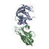

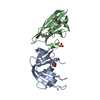
 Mass: 18.015 Da / Num. of mol.: 86 / Source method: isolated from a natural source / Formula: H2O
 Sample preparation
 / Beamline: 22-BM / Wavelength: 1 Å
 Processing