[English] 日本語
Yorodumi
- PDB-2p44: Complex of a camelid single-domain vhh antibody fragment with RNA... -

+
Open data


ID or keywords:

Loading...

-
Basic information

Entry
Database: PDB / ID: 2p44
Title
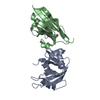
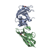
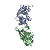
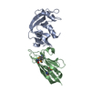
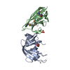
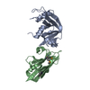
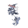
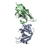
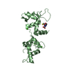
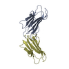
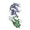
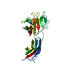
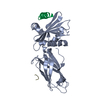
Complex of a camelid single-domain vhh antibody fragment with RNASE A at 1.8A resolution: SE5A-mono-1 crystal form with five se-met sites (M34, M51, F68M, M83, L86M) in vhh scaffold
Components
  • ANTIBODY CAB-RN05
  • Ribonuclease pancreaticPancreatic ribonuclease family
KeywordsHYDROLASE/IMMUNE SYSTEM / SEMET PHASING / CAMELID SINGLE-DOMAIN ANTIBODY / VHH / CAB-RN05 / RNASE A / YEAST SURFACE DISPLAY / HYDROLASE / HYDROLASE-IMMUNE SYSTEM COMPLEX
Function / homology
Function and homology information


pancreatic ribonuclease / ribonuclease A activity / RNA nuclease activity / nucleic acid binding / lyase activity / defense response to Gram-positive bacterium / extracellular region
Similarity search - Function
P-30 Protein / Ribonuclease A-like domain / Pancreatic ribonuclease / Ribonuclease A, active site / Ribonuclease A-domain / Ribonuclease A-like domain superfamily / Pancreatic ribonuclease / Pancreatic ribonuclease family signature. / Pancreatic ribonuclease / Immunoglobulins ...P-30 Protein / Ribonuclease A-like domain / Pancreatic ribonuclease / Ribonuclease A, active site / Ribonuclease A-domain / Ribonuclease A-like domain superfamily / Pancreatic ribonuclease / Pancreatic ribonuclease family signature. / Pancreatic ribonuclease / Immunoglobulins / Roll / Immunoglobulin-like / Sandwich / Mainly Beta / Alpha Beta
Similarity search - Domain/homology
Ribonuclease pancreatic
Similarity search - Component
Biological speciesCamelus dromedarius (Arabian camel)
Bos taurus (cattle)
MethodX-RAY DIFFRACTION / SYNCHROTRON / MAD / Resolution: 1.8 Å
AuthorsTereshko, V. / Uysal, S. / Koide, A. / Margalef, K. / Koide, S. / Kossiakoff, A.A.
CitationJournal: Protein Sci. / Year: 2008
Title: Toward chaperone-assisted crystallography: protein engineering enhancement of crystal packing and X-ray phasing capabilities of a camelid single-domain antibody (VHH) scaffold
Authors: Tereshko, V. / Uysal, S. / Koide, A. / Margalef, K. / Koide, S. / Kossiakoff, A.A.
History
DepositionMar 11, 2007Deposition site: RCSB / Processing site: RCSB
Revision 1.0Mar 11, 2008Provider: repository / Type: Initial release
Revision 1.1Jul 13, 2011Group: Advisory / Refinement description / Version format compliance
Remark 999 SEQUENCE AT THE TIME OF PROCESSING, THE SEQUENCE OF VHH ANTIBODY IS NOT AVAILABLE AT THE UNP ... SEQUENCE AT THE TIME OF PROCESSING, THE SEQUENCE OF VHH ANTIBODY IS NOT AVAILABLE AT THE UNP SEQUENCE DATABASE.

-
Structure visualization

Structure viewerMolecule:
MolmilJmol/JSmol

Downloads & links

-
Assembly

Deposited unit
A: Ribonuclease pancreatic
B: ANTIBODY CAB-RN05


Theoretical massNumber of molelcules
Total (without water)27,0492
Polymers27,0492
Non-polymers00
Water2,522140
1


  • Idetical with deposited unit
  • defined by author&software
TypeNameSymmetry operationNumber
identity operation1_555x,y,z1
Buried area1160 Å2
ΔGint-4.3 kcal/mol
Surface area12130 Å2
MethodPISA
Unit cell
Length a, b, c (Å)40.783, 54.765, 49.064
Angle α, β, γ (deg.)90.00, 109.57, 90.00
Int Tables number4
Space group name H-MP1211

-
Components

#1: Protein Ribonuclease pancreatic / Pancreatic ribonuclease family / RNase 1 / RNase A


Mass: 13708.326 Da / Num. of mol.: 1 / Source method: isolated from a natural source / Source: (natural) Bos taurus (cattle) / References: UniProt: P61823, EC: 3.1.27.5
#2: Protein ANTIBODY CAB-RN05


Mass: 13341.001 Da / Num. of mol.: 1
Source method: isolated from a genetically manipulated source
Source: (gene. exp.) Camelus dromedarius (Arabian camel) / Production host: Escherichia coli (E. coli)
#3: Water ChemComp-HOH / water / Water


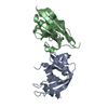
Mass: 18.015 Da / Num. of mol.: 140 / Source method: isolated from a natural source / Formula: H2O

-
Experimental details

-
Experiment

ExperimentMethod: X-RAY DIFFRACTION / Number of used crystals: 1

-
Sample preparation

CrystalDensity Matthews: 1.91 Å3/Da / Density % sol: 35.54 %
Crystal growTemperature: 295 K / Method: vapor diffusion / pH: 8.5
Details: HAMPTON RESEARCH INDEX SCREEN, SOLUTION #85: MGCL2, PEG 3350, TRIS BUFFER, PH 8.5, VAPOR DIFFUSION, temperature 295K

-
Data collection

DiffractionMean temperature: 100 K
Diffraction sourceSource: SYNCHROTRON / Site: APS / Beamline: 19-ID / Wavelength: 0.97955, 0.97934
DetectorType: SBC-3 / Detector: CCD / Date: Aug 1, 2001 / Details: MIRRORS
RadiationProtocol: MAD / Monochromatic (M) / Laue (L): M / Scattering type: x-ray
Radiation wavelength
IDWavelength (Å)Relative weight
10.979551
20.979341
ReflectionResolution: 1.8→20 Å / Num. all: 18982 / Num. obs: 18982 / % possible obs: 99.9 % / Observed criterion σ(F): 0 / Observed criterion σ(I): 0 / Redundancy: 3.3 % / Rmerge(I) obs: 0.095

-
Processing

Software
NameVersionClassification
REFMAC5.2.0005refinement
HKL-3000data collection
HKL-3000data reduction
HKL-3000data scaling
SOLVEphasing
RefinementMethod to determine structure: MAD / Resolution: 1.8→20 Å / Cor.coef. Fo:Fc: 0.951 / Cor.coef. Fo:Fc free: 0.921 / SU B: 6.662 / SU ML: 0.096 / TLS residual ADP flag: LIKELY RESIDUAL / Cross valid method: THROUGHOUT / σ(F): 0 / σ(I): 0 / ESU R: 0.157 / ESU R Free: 0.145 / Stereochemistry target values: MAXIMUM LIKELIHOOD / Details: HYDROGENS HAVE BEEN ADDED IN THE RIDING POSITIONS
RfactorNum. reflection% reflectionSelection details
Rfree0.2382 970 5.1 %RANDOM
Rwork0.19324 ---
all0.19544 18982 --
obs0.19544 18012 99.98 %-
Solvent computationIon probe radii: 0.8 Å / Shrinkage radii: 0.8 Å / VDW probe radii: 1.2 Å / Solvent model: BABINET MODEL WITH MASK
Displacement parametersBiso mean: 21.42 Å2
Baniso -1Baniso -2Baniso -3
1--0.16 Å20 Å2-1.09 Å2
2---1.3 Å20 Å2
3---0.73 Å2
Refinement stepCycle: LAST / Resolution: 1.8→20 Å
ProteinNucleic acidLigandSolventTotal
Num. atoms1840 0 0 140 1980
Refine LS restraints
Refine-IDTypeDev idealDev ideal targetNumber
X-RAY DIFFRACTIONr_bond_refined_d0.010.0191876
X-RAY DIFFRACTIONr_bond_other_d0.0010.021599
X-RAY DIFFRACTIONr_angle_refined_deg1.411.8922531
X-RAY DIFFRACTIONr_angle_other_deg0.7582.1533741
X-RAY DIFFRACTIONr_dihedral_angle_1_deg6.4285239
X-RAY DIFFRACTIONr_dihedral_angle_2_deg29.52324.28684
X-RAY DIFFRACTIONr_dihedral_angle_3_deg13.22615315
X-RAY DIFFRACTIONr_dihedral_angle_4_deg11.2191511
X-RAY DIFFRACTIONr_chiral_restr0.070.2268
X-RAY DIFFRACTIONr_gen_planes_refined0.0050.022130
X-RAY DIFFRACTIONr_gen_planes_other0.0010.02379
X-RAY DIFFRACTIONr_nbd_refined0.2110.2296
X-RAY DIFFRACTIONr_nbd_other0.190.21517
X-RAY DIFFRACTIONr_nbtor_refined0.1870.2917
X-RAY DIFFRACTIONr_nbtor_other0.0830.21031
X-RAY DIFFRACTIONr_xyhbond_nbd_refined0.1350.2101
X-RAY DIFFRACTIONr_xyhbond_nbd_other
X-RAY DIFFRACTIONr_metal_ion_refined
X-RAY DIFFRACTIONr_metal_ion_other
X-RAY DIFFRACTIONr_symmetry_vdw_refined0.1670.218
X-RAY DIFFRACTIONr_symmetry_vdw_other0.2510.253
X-RAY DIFFRACTIONr_symmetry_hbond_refined0.1070.213
X-RAY DIFFRACTIONr_symmetry_hbond_other
X-RAY DIFFRACTIONr_symmetry_metal_ion_refined
X-RAY DIFFRACTIONr_symmetry_metal_ion_other
X-RAY DIFFRACTIONr_mcbond_it1.2311.51481
X-RAY DIFFRACTIONr_mcbond_other0.2941.5506
X-RAY DIFFRACTIONr_mcangle_it1.41921897
X-RAY DIFFRACTIONr_scbond_it2.4193803
X-RAY DIFFRACTIONr_scangle_it3.0644.5634
X-RAY DIFFRACTIONr_rigid_bond_restr
X-RAY DIFFRACTIONr_sphericity_free
X-RAY DIFFRACTIONr_sphericity_bonded
LS refinement shellResolution: 1.8→1.846 Å / Total num. of bins used: 20
RfactorNum. reflection% reflection
Rfree0.262 78 -
Rwork0.2 1300 -
obs--99.86 %
Refinement TLS params.

Method: refined / Refine-ID: X-RAY DIFFRACTION

IDL112)L122)L132)L222)L232)L332)S11 (Å °)S12 (Å °)S13 (Å °)S21 (Å °)S22 (Å °)S23 (Å °)S31 (Å °)S32 (Å °)S33 (Å °)T112)T122)T132)T222)T232)T332)Origin x (Å)Origin y (Å)Origin z (Å)
12.4257-2.1275-0.07222.8917-0.49991.0681-0.4313-0.3089-0.04110.53920.3016-0.04680.00840.1530.12970.03930.06320.01490.02610.0134-0.059121.790634.032717.8347
21.5459-1.12840.45991.4598-0.50730.65150.01390.085-0.098-0.03530.03210.1433-0.027-0.0235-0.046-0.06520.00110.0085-0.04710.0014-0.03821.434352.01540.7391
Refinement TLS group
IDRefine-IDRefine TLS-IDAuth asym-IDAuth seq-ID
1X-RAY DIFFRACTION1A1 - 124
2X-RAY DIFFRACTION1A125 - 175
3X-RAY DIFFRACTION1B122 - 123
4X-RAY DIFFRACTION2B0 - 121
5X-RAY DIFFRACTION2A176 - 177
6X-RAY DIFFRACTION2B124 - 196
7X-RAY DIFFRACTION2A178 - 183
8X-RAY DIFFRACTION2B197 - 202

+
About Yorodumi

-
News

-
Feb 9, 2022. New format data for meta-information of EMDB entries

New format data for meta-information of EMDB entries

  • Version 3 of the EMDB header file is now the official format.
  • The previous official version 1.9 will be removed from the archive.

Related info.:EMDB header

External links:wwPDB to switch to version 3 of the EMDB data model

-
Aug 12, 2020. Covid-19 info

Covid-19 info

URL: https://pdbj.org/emnavi/covid19.php

New page: Covid-19 featured information page in EM Navigator.

Related info.:Covid-19 info / Mar 5, 2020. Novel coronavirus structure data

+
Mar 5, 2020. Novel coronavirus structure data

Novel coronavirus structure data

Related info.:Yorodumi Speices / Aug 12, 2020. Covid-19 info

External links:COVID-19 featured content - PDBj / Molecule of the Month (242):Coronavirus Proteases

+
Jan 31, 2019. EMDB accession codes are about to change! (news from PDBe EMDB page)

EMDB accession codes are about to change! (news from PDBe EMDB page)

  • The allocation of 4 digits for EMDB accession codes will soon come to an end. Whilst these codes will remain in use, new EMDB accession codes will include an additional digit and will expand incrementally as the available range of codes is exhausted. The current 4-digit format prefixed with “EMD-” (i.e. EMD-XXXX) will advance to a 5-digit format (i.e. EMD-XXXXX), and so on. It is currently estimated that the 4-digit codes will be depleted around Spring 2019, at which point the 5-digit format will come into force.
  • The EM Navigator/Yorodumi systems omit the EMD- prefix.

Related info.:Q: What is EMD? / ID/Accession-code notation in Yorodumi/EM Navigator

External links:EMDB Accession Codes are Changing Soon! / Contact to PDBj

+
Jul 12, 2017. Major update of PDB

Major update of PDB

  • wwPDB released updated PDB data conforming to the new PDBx/mmCIF dictionary.
  • This is a major update changing the version number from 4 to 5, and with Remediation, in which all the entries are updated.
  • In this update, many items about electron microscopy experimental information are reorganized (e.g. em_software).
  • Now, EM Navigator and Yorodumi are based on the updated data.

External links:wwPDB Remediation / Enriched Model Files Conforming to OneDep Data Standards Now Available in the PDB FTP Archive

-
Yorodumi

Thousand views of thousand structures

  • Yorodumi is a browser for structure data from EMDB, PDB, SASBDB, etc.
  • This page is also the successor to EM Navigator detail page, and also detail information page/front-end page for Omokage search.
  • The word "yorodu" (or yorozu) is an old Japanese word meaning "ten thousand". "mi" (miru) is to see.

Related info.:EMDB / PDB / SASBDB / Comparison of 3 databanks / Yorodumi Search / Aug 31, 2016. New EM Navigator & Yorodumi / Yorodumi Papers / Jmol/JSmol / Function and homology information / Changes in new EM Navigator and Yorodumi

Read more