 Movie
Movie Controller

[English] 日本語
 Yorodumi
- PDB-5e3q: Crystal structure of DapD in complex with succinyl-CoA from Coryn... -
+ Open data

- Basic information
| Entry | Database: PDB / ID: 5e3q | ||||||
|---|---|---|---|---|---|---|---|
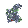
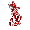
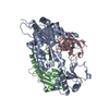
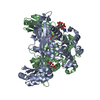
| Title | Crystal structure of DapD in complex with succinyl-CoA from Corynebacterium glutamicum | ||||||
 Components Components | 2,3,4,5-tetrahydropyridine-2,6-dicarboxylate N-succinyltransferase | ||||||
 Keywords Keywords | TRANSFERASE / Corynebacterium glutamicum / L-lysine | ||||||
| Function / homology |  Function and homology information Function and homology information2,3,4,5-tetrahydropyridine-2,6-dicarboxylate N-succinyltransferase / 2,3,4,5-tetrahydropyridine-2,6-dicarboxylate N-succinyltransferase activity / : / : / magnesium ion binding / cytoplasm Similarity search - Function | ||||||
| Biological species |  Corynebacterium glutamicum (bacteria) Corynebacterium glutamicum (bacteria) | ||||||
| Method |  X-RAY DIFFRACTION / SYNCHROTRON / MOLECULAR REPLACEMENT / Resolution: 1.8 Å X-RAY DIFFRACTION / SYNCHROTRON / MOLECULAR REPLACEMENT / Resolution: 1.8 Å | ||||||
 Authors Authors | Sagong, H.-Y. / Kim, K.-J. | ||||||
 Citation Citation | Journal: J.Agric.Food Chem. / Year: 2015 Title: Crystal Structure and Biochemical Characterization of Tetrahydrodipicolinate N-Succinyltransferase from Corynebacterium glutamicum. Authors: Sagong, H.Y. / Kim, K.J. | ||||||
| History |
|
- Structure visualization
| Structure viewer | Molecule: MolmilJmol/JSmol |
|---|
- Downloads & links
-Download
| PDBx/mmCIF format | 5e3q.cif.gz | 75.2 KB | Display | PDBx/mmCIF format |
|---|---|---|---|---|
| PDB format | pdb5e3q.ent.gz | 53 KB | Display | PDB format |
| PDBx/mmJSON format | 5e3q.json.gz | Tree view | PDBx/mmJSON format | |
| Others |  Other downloads Other downloads |
-Validation report
| Arichive directory | https://data.pdbj.org/pub/pdb/validation_reports/e3/5e3qftp://data.pdbj.org/pub/pdb/validation_reports/e3/5e3q | HTTPS FTP |
|---|
-Related structure data
| Related structure data |  5e3pC  5e3rC  3fsxS C: citing same article ( S: Starting model for refinement |
|---|---|
| Similar structure data |


-Links
 PDBj
PDBj


- Assembly
| Deposited unit | 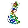
| |||||||||||||||
|---|---|---|---|---|---|---|---|---|---|---|---|---|---|---|---|---|
| 1 | 
| |||||||||||||||
| Unit cell |
| |||||||||||||||
| Components on special symmetry positions |
|
-Components
| #1: Protein | Mass: 31909.848 Da / Num. of mol.: 1 Source method: isolated from a genetically manipulated source Source: (gene. exp.) Corynebacterium glutamicum (strain ATCC 13032 / DSM 20300 / JCM 1318 / LMG 3730 / NCIMB 10025) (bacteria)Strain: ATCC 13032 / DSM 20300 / JCM 1318 / LMG 3730 / NCIMB 10025 Gene: dapD, Cgl1106, cg1256 / Plasmid: pET30a / Production host: References: UniProt: Q8NRE3, 2,3,4,5-tetrahydropyridine-2,6-dicarboxylate N-succinyltransferase |
|---|---|
| #2: Chemical | ChemComp-SCA /   Mass: 867.607 Da / Num. of mol.: 1 / Source method: obtained synthetically / Formula: C25H40N7O19P3S Mass: 867.607 Da / Num. of mol.: 1 / Source method: obtained synthetically / Formula: C25H40N7O19P3S |
| #3: Water | ChemComp-HOH /  Mass: 18.015 Da / Num. of mol.: 230 / Source method: isolated from a natural source / Formula: H2O Mass: 18.015 Da / Num. of mol.: 230 / Source method: isolated from a natural source / Formula: H2O |
-Experimental details
-Experiment
| Experiment | Method: X-RAY DIFFRACTION / Number of used crystals: 1 |
|---|
- Sample preparation
| Crystal | Density Matthews: 3.97 Å3/Da / Density % sol: 69.02 % |
|---|---|
| Crystal grow | Temperature: 293 K / Method: vapor diffusion, hanging drop / pH: 7 / Details: PEG 200, Li2SO4 |
-Data collection
| Diffraction | Mean temperature: 100 K |
|---|---|
| Diffraction source | Source: SYNCHROTRON / Site: PAL/PLS  / Beamline: 7A (6B, 6C1) / Wavelength: 0.97934 Å / Beamline: 7A (6B, 6C1) / Wavelength: 0.97934 Å |
| Detector | Type: ADSC QUANTUM 270 / Detector: CCD / Date: May 19, 2015 |
| Radiation | Monochromator: Double Crystal Monochromator / Protocol: SINGLE WAVELENGTH / Monochromatic (M) / Laue (L): M / Scattering type: x-ray |
| Radiation wavelength | Wavelength: 0.97934 Å / Relative weight: 1 |
| Reflection | Resolution: 1.8→94.11 Å / Num. obs: 41214 / % possible obs: 99.17 % / Redundancy: 10.5 % / Net I/σ(I): 61.68 |
- Processing
| Software |
| |||||||||||||||||||||||||||||||||||||||||||||||||||||||||||||||||||||||||||
|---|---|---|---|---|---|---|---|---|---|---|---|---|---|---|---|---|---|---|---|---|---|---|---|---|---|---|---|---|---|---|---|---|---|---|---|---|---|---|---|---|---|---|---|---|---|---|---|---|---|---|---|---|---|---|---|---|---|---|---|---|---|---|---|---|---|---|---|---|---|---|---|---|---|---|---|---|
| Refinement | Method to determine structure: MOLECULAR REPLACEMENT Starting model: 3FSX Resolution: 1.8→94.11 Å / Cor.coef. Fo:Fc: 0.963 / Cor.coef. Fo:Fc free: 0.951 / SU B: 1.455 / SU ML: 0.047 / Cross valid method: THROUGHOUT / σ(F): 0 / ESU R: 0.078 / ESU R Free: 0.082 / Stereochemistry target values: MAXIMUM LIKELIHOOD / Details: HYDROGENS HAVE BEEN ADDED IN THE RIDING POSITIONS
| |||||||||||||||||||||||||||||||||||||||||||||||||||||||||||||||||||||||||||
| Solvent computation | Ion probe radii: 0.8 Å / Shrinkage radii: 0.8 Å / VDW probe radii: 1.2 Å / Solvent model: MASK | |||||||||||||||||||||||||||||||||||||||||||||||||||||||||||||||||||||||||||
| Displacement parameters | Biso max: 82.86 Å2 / Biso mean: 23.175 Å2 / Biso min: 9.91 Å2
| |||||||||||||||||||||||||||||||||||||||||||||||||||||||||||||||||||||||||||
| Refinement step | Cycle: final / Resolution: 1.8→94.11 Å
| |||||||||||||||||||||||||||||||||||||||||||||||||||||||||||||||||||||||||||
| Refine LS restraints |
| |||||||||||||||||||||||||||||||||||||||||||||||||||||||||||||||||||||||||||
| LS refinement shell | Resolution: 1.799→1.845 Å / Total num. of bins used: 20
|
