 Movie
Movie Controller


+ Open data

- Basic information
| Entry | Database: PDB / ID: 3pod | ||||||
|---|---|---|---|---|---|---|---|
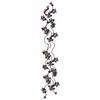
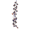
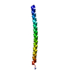
| Title | Crystal structure of MBL collagen-like peptide | ||||||
 Components Components | MBL collagen-like peptide | ||||||
 Keywords Keywords | UNKNOWN FUNCTION / Collagen peptide / complement proteins / lectin pathway of complement / MASP-1 CUB1 and CUB2 domains / Bloodstream | ||||||
| Method |  X-RAY DIFFRACTION / SYNCHROTRON / MOLECULAR REPLACEMENT / Resolution: 1.497 Å X-RAY DIFFRACTION / SYNCHROTRON / MOLECULAR REPLACEMENT / Resolution: 1.497 Å | ||||||
 Authors Authors | Gingras, A.R. / Moody, P.C.E. / Wallis, R. | ||||||
 Citation Citation | Journal: Structure / Year: 2011 Title: Structural Basis of Mannan-Binding Lectin Recognition by Its Associated Serine Protease MASP-1: Implications for Complement Activation. Authors: Gingras, A.R. / Girija, U.V. / Keeble, A.H. / Panchal, R. / Mitchell, D.A. / Moody, P.C. / Wallis, R. | ||||||
| History |
|
- Structure visualization
| Structure viewer | Molecule:  MolmilJmol/JSmol MolmilJmol/JSmol |
|---|
- Downloads & links
-Download
| PDBx/mmCIF format | 3pod.cif.gz | 36.5 KB | Display | PDBx/mmCIF format |
|---|---|---|---|---|
| PDB format | pdb3pod.ent.gz | 29 KB | Display | PDB format |
| PDBx/mmJSON format | 3pod.json.gz | Tree view | PDBx/mmJSON format | |
| Others |  Other downloads Other downloads |
-Validation report
| Arichive directory | https://data.pdbj.org/pub/pdb/validation_reports/po/3podftp://data.pdbj.org/pub/pdb/validation_reports/po/3pod | HTTPS FTP |
|---|
-Related structure data
| Related structure data |  3pobC 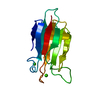 3poeC  3pofC 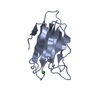 3pogC 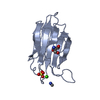 3poiC 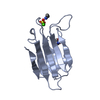 3pojC  3ponC 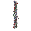 1cagS C: citing same article ( S: Starting model for refinement |
|---|---|
| Similar structure data |


-Links
 PDBj
PDBj- Assembly
| Deposited unit | 
| ||||||||
|---|---|---|---|---|---|---|---|---|---|
| 1 |
| ||||||||
| 2 | 
| ||||||||
| 3 | 
| ||||||||
| Unit cell |
|
-Components
| #1: Protein/peptide | Mass: 2212.419 Da / Num. of mol.: 3 / Source method: obtained synthetically / Details: peptide #2: Water | ChemComp-HOH / |  Mass: 18.015 Da / Num. of mol.: 94 / Source method: isolated from a natural source / Formula: H2O Mass: 18.015 Da / Num. of mol.: 94 / Source method: isolated from a natural source / Formula: H2O |
|---|
-Experimental details
-Experiment
| Experiment | Method: X-RAY DIFFRACTION / Number of used crystals: 1 |
|---|
- Sample preparation
| Crystal | Density Matthews: 1.93 Å3/Da / Density % sol: 36.22 % |
|---|---|
| Crystal grow | Temperature: 293 K / Method: vapor diffusion, sitting drop / pH: 6 Details: 1.6M ammonium sulfate, 100mM MES buffer, pH 6.0, VAPOR DIFFUSION, SITTING DROP, temperature 293K |
-Data collection
| Diffraction | Mean temperature: 100 K |
|---|---|
| Diffraction source | Source: SYNCHROTRON / Site: ESRF  / Beamline: ID14-2 / Wavelength: 0.933 Å / Beamline: ID14-2 / Wavelength: 0.933 Å |
| Detector | Type: ADSC QUANTUM 4 / Detector: CCD / Date: Apr 14, 2010 / Details: mirrors |
| Radiation | Protocol: SINGLE WAVELENGTH / Monochromatic (M) / Laue (L): M / Scattering type: x-ray |
| Radiation wavelength | Wavelength: 0.933 Å / Relative weight: 1 |
| Reflection | Resolution: 1.497→50 Å / Num. all: 8301 / Num. obs: 8299 / % possible obs: 98.8 % / Observed criterion σ(F): 0 / Observed criterion σ(I): 0 / Redundancy: 2.98 % / Rmerge(I) obs: 0.045 / Net I/σ(I): 25.94 |
| Reflection shell | Resolution: 1.497→1.59 Å / Redundancy: 2.95 % / Rmerge(I) obs: 0.152 / Mean I/σ(I) obs: 10.15 / Num. unique all: 1300 / % possible all: 96.9 |
- Processing
| Software |
| ||||||||||||||||||||||||||||||||||||||||||||||||||||||||||||||||||||||||||||||||
|---|---|---|---|---|---|---|---|---|---|---|---|---|---|---|---|---|---|---|---|---|---|---|---|---|---|---|---|---|---|---|---|---|---|---|---|---|---|---|---|---|---|---|---|---|---|---|---|---|---|---|---|---|---|---|---|---|---|---|---|---|---|---|---|---|---|---|---|---|---|---|---|---|---|---|---|---|---|---|---|---|---|
| Refinement | Method to determine structure: MOLECULAR REPLACEMENT Starting model: PDB ENTRY 1CAG Resolution: 1.497→22.71 Å / Cor.coef. Fo:Fc: 0.981 / Cor.coef. Fo:Fc free: 0.952 / SU B: 2.312 / SU ML: 0.039 / Cross valid method: THROUGHOUT / ESU R: 0.079 / ESU R Free: 0.078 / Stereochemistry target values: MAXIMUM LIKELIHOOD / Details: HYDROGENS HAVE BEEN ADDED IN THE RIDING POSITIONS
| ||||||||||||||||||||||||||||||||||||||||||||||||||||||||||||||||||||||||||||||||
| Solvent computation | Ion probe radii: 0.8 Å / Shrinkage radii: 0.8 Å / VDW probe radii: 1.4 Å / Solvent model: MASK | ||||||||||||||||||||||||||||||||||||||||||||||||||||||||||||||||||||||||||||||||
| Displacement parameters | Biso mean: 8.125 Å2
| ||||||||||||||||||||||||||||||||||||||||||||||||||||||||||||||||||||||||||||||||
| Refinement step | Cycle: LAST / Resolution: 1.497→22.71 Å
| ||||||||||||||||||||||||||||||||||||||||||||||||||||||||||||||||||||||||||||||||
| Refine LS restraints |
| ||||||||||||||||||||||||||||||||||||||||||||||||||||||||||||||||||||||||||||||||
| LS refinement shell | Resolution: 1.497→1.535 Å / Total num. of bins used: 20
|
