 Movie
Movie Controller

[English] 日本語
 Yorodumi
- PDB-3a1h: Crystal Structure Analysis of the Collagen-like Peptide, (PPG)4-O... -
+ Open data

- Basic information
| Entry | Database: PDB / ID: 3a1h | ||||||
|---|---|---|---|---|---|---|---|
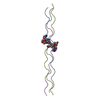
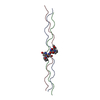
| Title | Crystal Structure Analysis of the Collagen-like Peptide, (PPG)4-OTG-(PPG)4 | ||||||
 Components Components | collagen-like peptide | ||||||
 Keywords Keywords | STRUCTURAL PROTEIN / collagen helix / host-guest peptide / twinned crystal | ||||||
| Method |  X-RAY DIFFRACTION / SYNCHROTRON / MOLECULAR REPLACEMENT / Resolution: 1.08 Å X-RAY DIFFRACTION / SYNCHROTRON / MOLECULAR REPLACEMENT / Resolution: 1.08 Å | ||||||
 Authors Authors | Okuyama, K. / Miyama, K. / Mizuno, K. / Bachinger, H.P. | ||||||
 Citation Citation | Journal: Biopolymers / Year: 2011 Title: Stabilization of triple-helical structures of collagen peptides containing a Hyp-Thr-Gly, Hyp-Val-Gly, or Hyp-Ser-Gly sequence. Authors: Okuyama, K. / Miyama, K. / Morimoto, T. / Masakiyo, K. / Mizuno, K. / Bachinger, H.P. | ||||||
| History |
|
- Structure visualization
| Structure viewer | Molecule:  MolmilJmol/JSmol MolmilJmol/JSmol |
|---|
- Downloads & links
-Download
| PDBx/mmCIF format | 3a1h.cif.gz | 60.4 KB | Display | PDBx/mmCIF format |
|---|---|---|---|---|
| PDB format | pdb3a1h.ent.gz | 47.5 KB | Display | PDB format |
| PDBx/mmJSON format | 3a1h.json.gz | Tree view | PDBx/mmJSON format | |
| Others |  Other downloads Other downloads |
-Validation report
| Arichive directory | https://data.pdbj.org/pub/pdb/validation_reports/a1/3a1hftp://data.pdbj.org/pub/pdb/validation_reports/a1/3a1h | HTTPS FTP |
|---|
-Related structure data
| Related structure data |  3a0mC 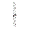 3admC 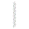 2cuoS S: Starting model for refinement C: citing same article ( |
|---|---|
| Similar structure data |


-Links
 PDBj
PDBj- Assembly
| Deposited unit | 
| ||||||||
|---|---|---|---|---|---|---|---|---|---|
| 1 | 
| ||||||||
| 2 | 
| ||||||||
| Unit cell |
|
-Components
| #1: Protein/peptide | Mass: 2299.536 Da / Num. of mol.: 6 / Source method: obtained synthetically / Details: THIS PEPTIDE WAS CHEMICALLY SYSTHESIZED. #2: Water | ChemComp-HOH / |  Mass: 18.015 Da / Num. of mol.: 222 / Source method: isolated from a natural source / Formula: H2O Mass: 18.015 Da / Num. of mol.: 222 / Source method: isolated from a natural source / Formula: H2OSequence details | THIS SEQUENCE ADOPTS A TRIPLE-HELICAL STRUCURE SIMILAR TO THE COLLAGEN-HELIX. | |
|---|
-Experimental details
-Experiment
| Experiment | Method: X-RAY DIFFRACTION / Number of used crystals: 1 |
|---|
- Sample preparation
| Crystal | Density Matthews: 1.99 Å3/Da / Density % sol: 38.54 % |
|---|---|
| Crystal grow | Temperature: 277 K / Method: vapor diffusion, sitting drop / pH: 5.9 Details: 12.5%(w/v) PEG 400, 0.1M MES buffer, pH 5.9, VAPOR DIFFUSION, SITTING DROP, temperature 277K |
-Data collection
| Diffraction | Mean temperature: 100 K |
|---|---|
| Diffraction source | Source: SYNCHROTRON / Site: SPring-8  / Beamline: BL41XU / Wavelength: 1 Å / Beamline: BL41XU / Wavelength: 1 Å |
| Detector | Type: ADSC QUANTUM 315 / Detector: CCD / Date: Jan 29, 2008 |
| Radiation | Monochromator: the rotated-inclined double crystal monochromator Protocol: SINGLE WAVELENGTH / Monochromatic (M) / Laue (L): M / Scattering type: x-ray |
| Radiation wavelength | Wavelength: 1 Å / Relative weight: 1 |
| Reflection twin | Operator: h,-k,-l / Fraction: 0.44 |
| Reflection | Resolution: 1.08→26.5 Å / Num. all: 47303 / Num. obs: 46090 / % possible obs: 97.4 % / Observed criterion σ(I): 3 / Redundancy: 3.26 % / Rmerge(I) obs: 0.076 |
| Reflection shell | Resolution: 1.08→1.12 Å / Redundancy: 3.33 % / Rmerge(I) obs: 0.304 / Mean I/σ(I) obs: 2.9 / Num. unique all: 4661 / % possible all: 99.1 |
- Processing
| Software |
| |||||||||||||||||||||||||||||||||
|---|---|---|---|---|---|---|---|---|---|---|---|---|---|---|---|---|---|---|---|---|---|---|---|---|---|---|---|---|---|---|---|---|---|---|
| Refinement | Method to determine structure: MOLECULAR REPLACEMENT Starting model: 2CUO Resolution: 1.08→26.5 Å / Num. parameters: 9778 / Num. restraintsaints: 12276 / Isotropic thermal model: anisotropic / Cross valid method: THROUGHOUT / σ(F): 0 / Stereochemistry target values: Engh & Huber Details: THE STRUCTURE WAS REFINED UNDER THE TWINNING OPERATOR (H,-K,-L) AND TWINNING FRACTION 0.44 USING THE TWINNED DATA.
| |||||||||||||||||||||||||||||||||
| Refine analyze | Num. disordered residues: 0 / Occupancy sum hydrogen: 0 / Occupancy sum non hydrogen: 1086 | |||||||||||||||||||||||||||||||||
| Refinement step | Cycle: LAST / Resolution: 1.08→26.5 Å
| |||||||||||||||||||||||||||||||||
| Refine LS restraints |
| |||||||||||||||||||||||||||||||||
| LS refinement shell |
|
