[English] 日本語
Yorodumi
- PDB-3pc8: X-ray crystal structure of the heterodimeric complex of XRCC1 and... -

+
Open data


ID or keywords:

Loading...

-
Basic information

Entry
Database: PDB / ID: 3pc8
Title
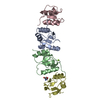
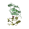
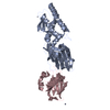
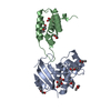
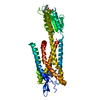
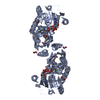
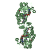
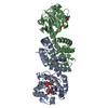
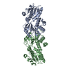
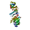
X-ray crystal structure of the heterodimeric complex of XRCC1 and DNA ligase III-alpha BRCT domains.
Components
  • DNA ligase 3
  • DNA repair protein XRCC1
KeywordsDNA BINDING PROTEIN/LIGASE / DNA repair / BRCT domain / Protein:protein interactions / DNA BINDING PROTEIN - LIGASE complex / DNA BINDING PROTEIN-LIGASE complex
Function / homology
Function and homology information


Resolution of AP sites via the single-nucleotide replacement pathway / HDR through MMEJ (alt-NHEJ) / DNA ligase III-XRCC1 complex / negative regulation of mitochondrial DNA replication / oxidized DNA binding / telomeric DNA-containing double minutes formation / ERCC4-ERCC1 complex / negative regulation of protection from non-homologous end joining at telomere / APEX1-Independent Resolution of AP Sites via the Single Nucleotide Replacement Pathway / base-excision repair, DNA ligation ...Resolution of AP sites via the single-nucleotide replacement pathway / HDR through MMEJ (alt-NHEJ) / DNA ligase III-XRCC1 complex / negative regulation of mitochondrial DNA replication / oxidized DNA binding / telomeric DNA-containing double minutes formation / ERCC4-ERCC1 complex / negative regulation of protection from non-homologous end joining at telomere / APEX1-Independent Resolution of AP Sites via the Single Nucleotide Replacement Pathway / base-excision repair, DNA ligation / ADP-D-ribose modification-dependent protein binding / Gap-filling DNA repair synthesis and ligation in TC-NER / negative regulation of protein ADP-ribosylation / DNA ligase activity / poly-ADP-D-ribose binding / DNA ligase (ATP) / positive regulation of single strand break repair / DNA ligase (ATP) activity / voluntary musculoskeletal movement / replication-born double-strand break repair via sister chromatid exchange / cerebellum morphogenesis / single strand break repair / DNA ligation / double-strand break repair via alternative nonhomologous end joining / HDR through MMEJ (alt-NHEJ) / lagging strand elongation / response to hydroperoxide / mitochondrial DNA repair / Resolution of AP sites via the single-nucleotide replacement pathway / DNA biosynthetic process / APEX1-Independent Resolution of AP Sites via the Single Nucleotide Replacement Pathway / site of DNA damage / base-excision repair, gap-filling / mitochondrion organization / Gap-filling DNA repair synthesis and ligation in GG-NER / response to organic substance / hippocampus development / double-strand break repair via homologous recombination / base-excision repair / double-strand break repair via nonhomologous end joining / Gap-filling DNA repair synthesis and ligation in TC-NER / double-strand break repair / chromosome, telomeric region / response to hypoxia / response to xenobiotic stimulus / cell cycle / cell division / DNA repair / chromatin / nucleolus / enzyme binding / mitochondrion / DNA binding / zinc ion binding / nucleoplasm / ATP binding / nucleus
Similarity search - Function
DNA ligase 3, BRCT domain / DNA ligase 3 BRCT domain / DNA-repair protein Xrcc1, N-terminal / XRCC1, first (central) BRCT domain / XRCC1 N terminal domain / DNA ligase, ATP-dependent / DNA ligase, ATP-dependent, N-terminal / DNA ligase, ATP-dependent, N-terminal domain superfamily / DNA ligase N terminus / ATP-dependent DNA ligase signature 2. ...DNA ligase 3, BRCT domain / DNA ligase 3 BRCT domain / DNA-repair protein Xrcc1, N-terminal / XRCC1, first (central) BRCT domain / XRCC1 N terminal domain / DNA ligase, ATP-dependent / DNA ligase, ATP-dependent, N-terminal / DNA ligase, ATP-dependent, N-terminal domain superfamily / DNA ligase N terminus / ATP-dependent DNA ligase signature 2. / ATP-dependent DNA ligase AMP-binding site. / DNA ligase, ATP-dependent, C-terminal / ATP dependent DNA ligase C terminal region / DNA ligase, ATP-dependent, conserved site / ATP-dependent DNA ligase family profile. / BRCT domain / DNA ligase, ATP-dependent, central / ATP dependent DNA ligase domain / Zinc finger poly(ADP-ribose) polymerase (PARP)-type signature. / Zinc finger, PARP-type superfamily / Poly(ADP-ribose) polymerase and DNA-Ligase Zn-finger region / Zinc finger poly(ADP-ribose) polymerase (PARP)-type profile. / Poly(ADP-ribose) polymerase and DNA-Ligase Zn-finger region / Zinc finger, PARP-type / BRCT domain, a BRCA1 C-terminus domain / BRCA1 C Terminus (BRCT) domain / breast cancer carboxy-terminal domain / BRCT domain profile. / BRCT domain / BRCT domain superfamily / Galactose-binding-like domain superfamily / Nucleic acid-binding, OB-fold / Rossmann fold / 3-Layer(aba) Sandwich / Alpha Beta
Similarity search - Domain/homology
DNA ligase 3 / DNA repair protein XRCC1
Similarity search - Component
Biological speciesMus musculus (house mouse)
Homo sapiens (human)
MethodX-RAY DIFFRACTION / MOLECULAR REPLACEMENT / Resolution: 2.31 Å
AuthorsCuneo, M.J. / Krahn, J.M. / London, R.E.
CitationJournal: Nucleic Acids Res. / Year: 2011
Title: The structural basis for partitioning of the XRCC1/DNA ligase III-{alpha} BRCT-mediated dimer complexes.
Authors: Cuneo, M.J. / Gabel, S.A. / Krahn, J.M. / Ricker, M.A. / London, R.E.
History
DepositionOct 21, 2010Deposition site: RCSB / Processing site: RCSB
Revision 1.0Jun 15, 2011Provider: repository / Type: Initial release
Revision 1.1Jul 13, 2011Group: Version format compliance
Revision 1.2Nov 2, 2011Group: Database references
Revision 1.3Feb 21, 2024Group: Data collection / Database references / Derived calculations
Category: chem_comp_atom / chem_comp_bond ...chem_comp_atom / chem_comp_bond / database_2 / pdbx_struct_conn_angle / struct_conn / struct_ref_seq_dif / struct_site
Item: _database_2.pdbx_DOI / _database_2.pdbx_database_accession ..._database_2.pdbx_DOI / _database_2.pdbx_database_accession / _pdbx_struct_conn_angle.ptnr1_auth_seq_id / _pdbx_struct_conn_angle.ptnr3_auth_seq_id / _pdbx_struct_conn_angle.value / _struct_conn.pdbx_dist_value / _struct_conn.ptnr2_auth_seq_id / _struct_ref_seq_dif.details / _struct_site.pdbx_auth_asym_id / _struct_site.pdbx_auth_comp_id / _struct_site.pdbx_auth_seq_id

-
Structure visualization

Structure viewerMolecule:
MolmilJmol/JSmol

Downloads & links

-
Assembly

Deposited unit
A: DNA repair protein XRCC1
B: DNA repair protein XRCC1
C: DNA ligase 3
D: DNA ligase 3
hetero molecules


Theoretical massNumber of molelcules
Total (without water)43,5606
Polymers43,4134
Non-polymers1462
Water3,747208
1


  • Idetical with deposited unit
  • defined by author
TypeNameSymmetry operationNumber
identity operation1_555x,y,z1
2
A: DNA repair protein XRCC1
C: DNA ligase 3


Theoretical massNumber of molelcules
Total (without water)21,7072
Polymers21,7072
Non-polymers00
Water362
TypeNameSymmetry operationNumber
identity operation1_555x,y,z1
Buried area1290 Å2
ΔGint-1 kcal/mol
Surface area10080 Å2
MethodPISA
3
B: DNA repair protein XRCC1
D: DNA ligase 3
hetero molecules


Theoretical massNumber of molelcules
Total (without water)21,8534
Polymers21,7072
Non-polymers1462
Water362
TypeNameSymmetry operationNumber
identity operation1_555x,y,z1
Buried area1260 Å2
ΔGint-9 kcal/mol
Surface area9740 Å2
MethodPISA
Unit cell
Length a, b, c (Å)66.320, 163.480, 36.740
Angle α, β, γ (deg.)90.000, 90.000, 90.000
Int Tables number18
Space group name H-MP21212
Components on special symmetry positions
IDModelComponents
11A-21-

HOH

Noncrystallographic symmetry (NCS)NCS domain:
IDEns-IDDetails
11A
21B
12A
22B
13A
23B
14C
24D
15C
25D

NCS domain segments:
Dom-IDComponent-IDEns-IDSelection detailsAuth asym-IDAuth seq-ID
111chain A and (backbone or name CB) and (resseq 540:588 or resseq 599:626)A0
121chain A and (backbone or name CB) and (resseq 540:588 or resseq 599:626)A0
211chain B and (backbone or name CB) and (resseq 540:588 or resseq 599:626)B0
221chain B and (backbone or name CB) and (resseq 540:588 or resseq 599:626)B0
112chain A and (backbone or name CB) and (resseq 593:598 or resseq 627:630)A0
122chain A and (backbone or name CB) and (resseq 593:598 or resseq 627:630)A0
212chain B and (backbone or name CB) and (resseq 593:598 or resseq 627:630)B0
222chain B and (backbone or name CB) and (resseq 593:598 or resseq 627:630)B0
113chain A and resseq 589:592A589 - 592
213chain B and resseq 589:592B589 - 592
114chain C and (backbone or name CB) and (resseq 848:881 or resseq 888:890)C0
124chain C and (backbone or name CB) and (resseq 848:881 or resseq 888:890)C0
214chain D and (backbone or name CB) and (resseq 848:881 or resseq 888:890)D0
224chain D and (backbone or name CB) and (resseq 848:881 or resseq 888:890)D0
115chain C and (backbone or name CB) and (resseq 902:918)C0
215chain D and (backbone or name CB) and (resseq 902:918)D0

NCS ensembles :
ID
1
2
3
4
5

-
Components

#1: Protein DNA repair protein XRCC1 / / X-ray repair cross-complementing protein 1


Mass: 11753.289 Da / Num. of mol.: 2 / Fragment: unp residues 534-631
Source method: isolated from a genetically manipulated source
Source: (gene. exp.) Mus musculus (house mouse) / Gene: Xrcc-1, Xrcc1 / Plasmid: petDuet / Production host: Escherichia coli (E. coli) / Strain (production host): BL21-DE3-RIL / References: UniProt: Q60596
#2: Protein DNA ligase 3 / / DNA ligase III / Polydeoxyribonucleotide synthase [ATP] 3


Mass: 9953.292 Da / Num. of mol.: 2 / Fragment: unp residues 924-1008 / Mutation: C842S, C921S
Source method: isolated from a genetically manipulated source
Source: (gene. exp.) Homo sapiens (human) / Gene: LIG3 / Plasmid: pColaDuet / Production host: Escherichia coli (E. coli) / Strain (production host): BL21-DE3-RIL / References: UniProt: P49916, DNA ligase (ATP)
#3: Chemical ChemComp-MG / MAGNESIUM ION


Mass: 24.305 Da / Num. of mol.: 1 / Source method: obtained synthetically / Formula: Mg
#4: Chemical ChemComp-TRS / 2-AMINO-2-HYDROXYMETHYL-PROPANE-1,3-DIOL / TRIS BUFFER / Tris


Mass: 122.143 Da / Num. of mol.: 1 / Source method: obtained synthetically / Formula: C4H12NO3 / Comment: pH buffer*YM
#5: Water ChemComp-HOH / water / Water


Mass: 18.015 Da / Num. of mol.: 208 / Source method: isolated from a natural source / Formula: H2O

-
Experimental details

-
Experiment

ExperimentMethod: X-RAY DIFFRACTION / Number of used crystals: 1

-
Sample preparation

CrystalDensity Matthews: 2.29 Å3/Da / Density % sol: 46.38 %
Crystal growTemperature: 277 K / Method: vapor diffusion, hanging drop / pH: 8.5
Details: 30% PEG 4000, 0.1M Tris, and 0.2 M MgCl2 , pH 8.5, VAPOR DIFFUSION, HANGING DROP, temperature 277K

-
Data collection

DiffractionMean temperature: 100 K
Diffraction sourceSource: ROTATING ANODE / Type: RIGAKU MICROMAX-007 HF / Wavelength: 1.5418 Å
DetectorType: RIGAKU SATURN 92 / Detector: CCD / Date: Sep 25, 2010
RadiationMonochromator: Mirrors / Protocol: SINGLE WAVELENGTH / Monochromatic (M) / Laue (L): M / Scattering type: x-ray
Radiation wavelengthWavelength: 1.5418 Å / Relative weight: 1
ReflectionResolution: 2.31→35 Å / Num. all: 17621 / Num. obs: 17621 / % possible obs: 96 % / Observed criterion σ(F): 1 / Observed criterion σ(I): -3 / Biso Wilson estimate: 39.42 Å2 / Rmerge(I) obs: 0.05 / Net I/σ(I): 26.14
Reflection shell

Diffraction-ID: 1

Resolution (Å)Highest resolution (Å)Rmerge(I) obsMean I/σ(I) obsNum. measured obsNum. unique obs% possible all
2.31-2.370.2446.45916109483.2
2.37-2.430.1969.48492127899
2.43-2.50.17110.987411257100
2.5-2.580.13712.986131227100
2.58-2.660.13313.88304119399.9
2.66-2.760.114168009114899.8
2.76-2.860.08819.578621123100
2.86-2.980.07122.974931066100
2.98-3.110.05826.973371050100
3.11-3.260.05130.76862982100
3.26-3.440.05431.2629294098.1
3.44-3.650.05234.4495378887.1
3.65-3.90.04636.5267454664.6
3.9-4.210.03543435774293.2
4.21-4.610.02951.95005737100
4.61-5.160.02851.84651678100
5.16-5.960.02849.44058599100
5.96-7.30.02649.23450519100
7.3-10.320.02854.62597408100
10.320.0354.3135124695

-
Processing

Software
NameVersionClassificationNB
XSCALEdata processing
PHENIX1.6.4_486refinement
PDB_EXTRACT3.1data extraction
StructureStudiodata collection
XDSdata reduction
XSCALEdata scaling
PHASERphasing
RefinementMethod to determine structure: MOLECULAR REPLACEMENT / Resolution: 2.31→33.511 Å / Occupancy max: 1 / Occupancy min: 0.02 / FOM work R set: 0.8403 / SU ML: 0.29 / σ(F): 1.99 / Phase error: 22.13 / Stereochemistry target values: ML
RfactorNum. reflection% reflectionSelection details
Rfree0.2465 880 5 %random
Rwork0.1973 ---
obs0.1998 17611 95.92 %-
all-17611 --
Solvent computationShrinkage radii: 0.83 Å / VDW probe radii: 1.1 Å / Solvent model: FLAT BULK SOLVENT MODEL / Bsol: 41.675 Å2 / ksol: 0.314 e/Å3
Displacement parametersBiso max: 132.67 Å2 / Biso mean: 35.917 Å2 / Biso min: 9.49 Å2
Baniso -1Baniso -2Baniso -3
1-12.9745 Å2-0 Å2-0 Å2
2---10.1732 Å2-0 Å2
3----2.8013 Å2
Refinement stepCycle: LAST / Resolution: 2.31→33.511 Å
ProteinNucleic acidLigandSolventTotal
Num. atoms2830 0 9 208 3047
Refine LS restraints
Refine-IDTypeDev idealNumber
X-RAY DIFFRACTIONf_bond_d0.0043049
X-RAY DIFFRACTIONf_angle_d0.7214135
X-RAY DIFFRACTIONf_chiral_restr0.053428
X-RAY DIFFRACTIONf_plane_restr0.003550
X-RAY DIFFRACTIONf_dihedral_angle_d12.2721125
Refine LS restraints NCS
Ens-IDDom-IDAuth asym-IDNumberRefine-IDTypeRms dev position (Å)
11A370X-RAY DIFFRACTIONPOSITIONAL0.075
12B370X-RAY DIFFRACTIONPOSITIONAL0.075
21A45X-RAY DIFFRACTIONPOSITIONAL0.092
22B45X-RAY DIFFRACTIONPOSITIONAL0.092
31A34X-RAY DIFFRACTIONPOSITIONAL0.056
32B34X-RAY DIFFRACTIONPOSITIONAL0.056
41C178X-RAY DIFFRACTIONPOSITIONAL0.09
42D178X-RAY DIFFRACTIONPOSITIONAL0.09
51C85X-RAY DIFFRACTIONPOSITIONAL0.071
52D85X-RAY DIFFRACTIONPOSITIONAL0.071
LS refinement shell

Refine-ID: X-RAY DIFFRACTION / Total num. of bins used: 6

Resolution (Å)Rfactor RfreeNum. reflection RfreeRfactor RworkNum. reflection RworkNum. reflection all% reflection obs (%)
2.3071-2.45160.30081360.24472621275792
2.4516-2.64080.31411500.221828492999100
2.6408-2.90650.26391500.217728482998100
2.9065-3.32670.24261530.203228963049100
3.3267-4.190.26271290.20962453258284
4.19-33.51410.20021620.161230643226100
Refinement TLS params.

Method: refined / Refine-ID: X-RAY DIFFRACTION

IDL112)L122)L132)L222)L232)L332)S11 (Å °)S12 (Å °)S13 (Å °)S21 (Å °)S22 (Å °)S23 (Å °)S31 (Å °)S32 (Å °)S33 (Å °)T112)T122)T132)T222)T232)T332)Origin x (Å)Origin y (Å)Origin z (Å)
11.517-0.7995-0.08593.99391.15243.6521-0.00590.0086-0.14240.0388-0.01550.25050.043-0.051300.0264-0.03130.0210.10010.02450.0791-16.04855.2734-2.3471
21.40480.61410.91426.43190.73963.4038-0.01440.11340.1372-0.1134-0.01820.1727-0.0640.0771-0.00010.10670.02450.00280.14080.02050.1195-16.650129.5525-4.5323
33.98950.9275-0.53063.4252-0.88261.41380.1208-0.0371-0.15140.1143-0.1041-0.26440.13640.1829-0.01210.0375-0.0098-0.03810.0517-0.01960.0448-7.5565-17.8732-3.6406
44.7205-0.80340.90043.4441.54751.17080.04890.48920.3034-0.4284-0.0143-0.4709-0.21730.39030.00010.17460.00210.02360.26840.05110.2314-9.449553.3348-2.9753
Refinement TLS group
IDRefine-IDRefine TLS-IDSelection detailsAuth asym-IDAuth seq-ID
1X-RAY DIFFRACTION1chain AA534 - 630
2X-RAY DIFFRACTION2chain B or (chain X and resid 1) or (chain S and (resid 170:172 or resid 72 or resid 215))B1
3X-RAY DIFFRACTION3chain CC846 - 922
4X-RAY DIFFRACTION4chain DD845 - 922

+
About Yorodumi

-
News

-
Feb 9, 2022. New format data for meta-information of EMDB entries

New format data for meta-information of EMDB entries

  • Version 3 of the EMDB header file is now the official format.
  • The previous official version 1.9 will be removed from the archive.

Related info.:EMDB header

External links:wwPDB to switch to version 3 of the EMDB data model

-
Aug 12, 2020. Covid-19 info

Covid-19 info

URL: https://pdbj.org/emnavi/covid19.php

New page: Covid-19 featured information page in EM Navigator.

Related info.:Covid-19 info / Mar 5, 2020. Novel coronavirus structure data

+
Mar 5, 2020. Novel coronavirus structure data

Novel coronavirus structure data

Related info.:Yorodumi Speices / Aug 12, 2020. Covid-19 info

External links:COVID-19 featured content - PDBj / Molecule of the Month (242):Coronavirus Proteases

+
Jan 31, 2019. EMDB accession codes are about to change! (news from PDBe EMDB page)

EMDB accession codes are about to change! (news from PDBe EMDB page)

  • The allocation of 4 digits for EMDB accession codes will soon come to an end. Whilst these codes will remain in use, new EMDB accession codes will include an additional digit and will expand incrementally as the available range of codes is exhausted. The current 4-digit format prefixed with “EMD-” (i.e. EMD-XXXX) will advance to a 5-digit format (i.e. EMD-XXXXX), and so on. It is currently estimated that the 4-digit codes will be depleted around Spring 2019, at which point the 5-digit format will come into force.
  • The EM Navigator/Yorodumi systems omit the EMD- prefix.

Related info.:Q: What is EMD? / ID/Accession-code notation in Yorodumi/EM Navigator

External links:EMDB Accession Codes are Changing Soon! / Contact to PDBj

+
Jul 12, 2017. Major update of PDB

Major update of PDB

  • wwPDB released updated PDB data conforming to the new PDBx/mmCIF dictionary.
  • This is a major update changing the version number from 4 to 5, and with Remediation, in which all the entries are updated.
  • In this update, many items about electron microscopy experimental information are reorganized (e.g. em_software).
  • Now, EM Navigator and Yorodumi are based on the updated data.

External links:wwPDB Remediation / Enriched Model Files Conforming to OneDep Data Standards Now Available in the PDB FTP Archive

-
Yorodumi

Thousand views of thousand structures

  • Yorodumi is a browser for structure data from EMDB, PDB, SASBDB, etc.
  • This page is also the successor to EM Navigator detail page, and also detail information page/front-end page for Omokage search.
  • The word "yorodu" (or yorozu) is an old Japanese word meaning "ten thousand". "mi" (miru) is to see.

Related info.:EMDB / PDB / SASBDB / Comparison of 3 databanks / Yorodumi Search / Aug 31, 2016. New EM Navigator & Yorodumi / Yorodumi Papers / Jmol/JSmol / Function and homology information / Changes in new EM Navigator and Yorodumi

Read more