- PDB-3pc6: X-ray crystal structure of the second XRCC1 BRCT domain. -
+
Open data
ID or keywords:
Loading...
-
Basic information
Entry
Database: PDB / ID: 3pc6
Title
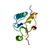
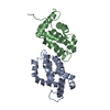
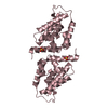
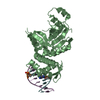
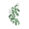
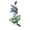
X-ray crystal structure of the second XRCC1 BRCT domain.
Components
DNA repair protein XRCC1
Keywords
DNA BINDING PROTEIN / DNA repair / BRCT domain / Protein:protein interactions / DNA ligase III-alpha BRCT2 domain
Function / homology
Function and homology information
Resolution of AP sites via the single-nucleotide replacement pathway / HDR through MMEJ (alt-NHEJ) / oxidized DNA binding / APEX1-Independent Resolution of AP Sites via the Single Nucleotide Replacement Pathway / telomeric DNA-containing double minutes formation / ERCC4-ERCC1 complex / negative regulation of protection from non-homologous end joining at telomere / ADP-D-ribose modification-dependent protein binding / Gap-filling DNA repair synthesis and ligation in TC-NER / poly-ADP-D-ribose binding ...Resolution of AP sites via the single-nucleotide replacement pathway / HDR through MMEJ (alt-NHEJ) / oxidized DNA binding / APEX1-Independent Resolution of AP Sites via the Single Nucleotide Replacement Pathway / telomeric DNA-containing double minutes formation / ERCC4-ERCC1 complex / negative regulation of protection from non-homologous end joining at telomere / ADP-D-ribose modification-dependent protein binding / Gap-filling DNA repair synthesis and ligation in TC-NER / poly-ADP-D-ribose binding / regulation of base-excision repair / single-strand break-containing DNA binding / single strand break repair / response to hydroperoxide / site of DNA damage / hippocampus development / base-excision repair / double-strand break repair via nonhomologous end joining / double-strand break repair / chromosome, telomeric region / DNA repair / nucleolus / chromatin / enzyme binding / nucleoplasm / nucleus Similarity search - Function
DNA-repair protein Xrcc1, N-terminal / XRCC1, first (central) BRCT domain / XRCC1 N terminal domain / BRCT domain / BRCT domain / BRCA1 C Terminus (BRCT) domain / breast cancer carboxy-terminal domain / BRCT domain profile. / BRCT domain / BRCT domain superfamily ...DNA-repair protein Xrcc1, N-terminal / XRCC1, first (central) BRCT domain / XRCC1 N terminal domain / BRCT domain / BRCT domain / BRCA1 C Terminus (BRCT) domain / breast cancer carboxy-terminal domain / BRCT domain profile. / BRCT domain / BRCT domain superfamily / Galactose-binding-like domain superfamily / Rossmann fold / 3-Layer(aba) Sandwich / Alpha Beta Similarity search - Domain/homology
In the structure databanks used in Yorodumi, some data are registered as the other names, "COVID-19 virus" and "2019-nCoV". Here are the details of the virus and the list of structure data.
Jan 31, 2019. EMDB accession codes are about to change! (news from PDBe EMDB page)
EMDB accession codes are about to change! (news from PDBe EMDB page)
The allocation of 4 digits for EMDB accession codes will soon come to an end. Whilst these codes will remain in use, new EMDB accession codes will include an additional digit and will expand incrementally as the available range of codes is exhausted. The current 4-digit format prefixed with “EMD-” (i.e. EMD-XXXX) will advance to a 5-digit format (i.e. EMD-XXXXX), and so on. It is currently estimated that the 4-digit codes will be depleted around Spring 2019, at which point the 5-digit format will come into force.
The EM Navigator/Yorodumi systems omit the EMD- prefix.
Related info.:Q: What is EMD? / ID/Accession-code notation in Yorodumi/EM Navigator
Yorodumi is a browser for structure data from EMDB, PDB, SASBDB, etc.
This page is also the successor to EM Navigator detail page, and also detail information page/front-end page for Omokage search.
The word "yorodu" (or yorozu) is an old Japanese word meaning "ten thousand". "mi" (miru) is to see.
Related info.:EMDB / PDB / SASBDB / Comparison of 3 databanks / Yorodumi Search / Aug 31, 2016. New EM Navigator & Yorodumi / Yorodumi Papers / Jmol/JSmol / Function and homology information / Changes in new EM Navigator and Yorodumi
 Movie
Movie Controller


 Open data
 Basic information
 Components
 Keywords
 Function and homology information
 X-RAY DIFFRACTION /
  Authors
 Citation
 Structure visualization
 Downloads & links
 Other downloads


 PDBj
PDBj

 Assembly
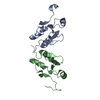

 Mass: 18.015 Da / Num. of mol.: 182 / Source method: isolated from a natural source / Formula: H2O
 Sample preparation
 / Beamline: 22-ID / Wavelength: 1 Å
 Processing