 Movie
Movie Controller


+ Open data

- Basic information
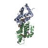
| Entry | Database: PDB / ID: 4m0s | ||||||
|---|---|---|---|---|---|---|---|
| Title | Crystal structure of Vaccinia virus protein A46 | ||||||
 Components Components | Toll/IL1-receptor signalling interference protein A46 | ||||||
 Keywords Keywords | VIRAL PROTEIN / BCl 2 fold / Toll/IL1-receptor signalling interference protein / protein binding | ||||||
| Function / homology | dsDNA poxvirus / Poxvirus Bcl-2-like / Poxvirus domain superfamily / Poxvirus Bcl-2-like proteins / Apoptosis Regulator Bcl-x / Orthogonal Bundle / Mainly Alpha / Putative 27.6k protein / Putative 27.6k protein Function and homology information Function and homology information | ||||||
| Biological species |  Vaccinia virus Vaccinia virus | ||||||
| Method |  X-RAY DIFFRACTION / SYNCHROTRON / MOLECULAR REPLACEMENT / Resolution: 2.58 Å X-RAY DIFFRACTION / SYNCHROTRON / MOLECULAR REPLACEMENT / Resolution: 2.58 Å | ||||||
 Authors Authors | Choe, J.W. / Kim, Y.W. | ||||||
 Citation Citation | Journal: To be Published Title: Crystal structure of Vaccinia virus protein A46 Authors: Choe, J.W. / Kim, Y.W. | ||||||
| History |
|
- Structure visualization
| Structure viewer | Molecule: MolmilJmol/JSmol |
|---|
- Downloads & links
-Download
| PDBx/mmCIF format | 4m0s.cif.gz | 130.9 KB | Display | PDBx/mmCIF format |
|---|---|---|---|---|
| PDB format | pdb4m0s.ent.gz | 105.8 KB | Display | PDB format |
| PDBx/mmJSON format | 4m0s.json.gz | Tree view | PDBx/mmJSON format | |
| Others |  Other downloads Other downloads |
-Validation report
| Arichive directory | https://data.pdbj.org/pub/pdb/validation_reports/m0/4m0sftp://data.pdbj.org/pub/pdb/validation_reports/m0/4m0s | HTTPS FTP |
|---|
-Related structure data
| Similar structure data |
|---|


-Links
 PDBj
PDBj- Assembly
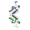
| Deposited unit | 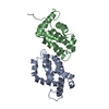
| ||||||||
|---|---|---|---|---|---|---|---|---|---|
| 1 |
| ||||||||
| Unit cell |
|
-Components
| #1: Protein | Mass: 17082.533 Da / Num. of mol.: 2 / Fragment: UNP RESIDUES 79-224 Source method: isolated from a genetically manipulated source Source: (gene. exp.) Vaccinia virus / Strain: Ankara / Gene: CVA180 / Production host:  #2: Water | ChemComp-HOH / |  Mass: 18.015 Da / Num. of mol.: 113 / Source method: isolated from a natural source / Formula: H2O Mass: 18.015 Da / Num. of mol.: 113 / Source method: isolated from a natural source / Formula: H2O |
|---|
-Experimental details
-Experiment
| Experiment | Method: X-RAY DIFFRACTION / Number of used crystals: 1 |
|---|
- Sample preparation
| Crystal grow | Temperature: 277.15 K / Method: vapor diffusion, hanging drop / pH: 7 Details: 1.3M sodium formate, 0.1M Tris pH 7.0 , VAPOR DIFFUSION, HANGING DROP, temperature 277.15K |
|---|
-Data collection
| Diffraction source | Source: SYNCHROTRON / Site: PAL/PLS  / Beamline: 7A (6B, 6C1) / Wavelength: 1 Å / Beamline: 7A (6B, 6C1) / Wavelength: 1 Å |
|---|---|
| Detector | Type: ADSC QUANTUM 210 / Detector: CCD |
| Radiation | Protocol: SINGLE WAVELENGTH / Monochromatic (M) / Laue (L): M / Scattering type: x-ray |
| Radiation wavelength | Wavelength: 1 Å / Relative weight: 1 |
| Reflection | Resolution: 2.58→26.79 Å / Num. obs: 29192 |
- Processing
| Software |
| ||||||||||||||||||||||||||||||||||||||||||||||||||||||||||||||||||||||||||||||||||||||||||||||||||||||||||||||||||||||||||||||||||||||||||||||||||||||||||||||||||||||||||||||||||||||
|---|---|---|---|---|---|---|---|---|---|---|---|---|---|---|---|---|---|---|---|---|---|---|---|---|---|---|---|---|---|---|---|---|---|---|---|---|---|---|---|---|---|---|---|---|---|---|---|---|---|---|---|---|---|---|---|---|---|---|---|---|---|---|---|---|---|---|---|---|---|---|---|---|---|---|---|---|---|---|---|---|---|---|---|---|---|---|---|---|---|---|---|---|---|---|---|---|---|---|---|---|---|---|---|---|---|---|---|---|---|---|---|---|---|---|---|---|---|---|---|---|---|---|---|---|---|---|---|---|---|---|---|---|---|---|---|---|---|---|---|---|---|---|---|---|---|---|---|---|---|---|---|---|---|---|---|---|---|---|---|---|---|---|---|---|---|---|---|---|---|---|---|---|---|---|---|---|---|---|---|---|---|---|---|
| Refinement | Method to determine structure: MOLECULAR REPLACEMENT / Resolution: 2.58→26.79 Å / Cor.coef. Fo:Fc: 0.955 / Cor.coef. Fo:Fc free: 0.938 / SU B: 13.577 / SU ML: 0.136 / Cross valid method: THROUGHOUT / ESU R: 0.191 / ESU R Free: 0.178 / Details: HYDROGENS HAVE BEEN ADDED IN THE RIDING POSITIONS
| ||||||||||||||||||||||||||||||||||||||||||||||||||||||||||||||||||||||||||||||||||||||||||||||||||||||||||||||||||||||||||||||||||||||||||||||||||||||||||||||||||||||||||||||||||||||
| Solvent computation | Ion probe radii: 0.8 Å / Shrinkage radii: 0.8 Å / VDW probe radii: 1.2 Å / Solvent model: MASK | ||||||||||||||||||||||||||||||||||||||||||||||||||||||||||||||||||||||||||||||||||||||||||||||||||||||||||||||||||||||||||||||||||||||||||||||||||||||||||||||||||||||||||||||||||||||
| Displacement parameters | Biso mean: 73.987 Å2
| ||||||||||||||||||||||||||||||||||||||||||||||||||||||||||||||||||||||||||||||||||||||||||||||||||||||||||||||||||||||||||||||||||||||||||||||||||||||||||||||||||||||||||||||||||||||
| Refinement step | Cycle: LAST / Resolution: 2.58→26.79 Å
| ||||||||||||||||||||||||||||||||||||||||||||||||||||||||||||||||||||||||||||||||||||||||||||||||||||||||||||||||||||||||||||||||||||||||||||||||||||||||||||||||||||||||||||||||||||||
| Refine LS restraints |
|