 Movie
Movie Controller


+ Open data

- Basic information
| Entry | Database: PDB / ID: 4lqk | ||||||
|---|---|---|---|---|---|---|---|
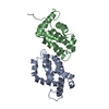
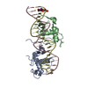
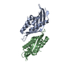
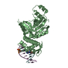
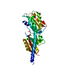
| Title | Structure of the vaccinia virus NF- B antagonist A46 | ||||||
 Components Components | Protein A46 | ||||||
 Keywords Keywords | VIRAL PROTEIN / Bcl-2-like fold | ||||||
| Function / homology |  Function and homology information Function and homology informationextrinsic component of cytoplasmic side of plasma membrane / protein sequestering activity / symbiont-mediated suppression of host toll-like receptor signaling pathway / symbiont-mediated suppression of host cytoplasmic pattern recognition receptor signaling pathway via inhibition of IRF3 activity / symbiont-mediated suppression of host NF-kappaB cascade Similarity search - Function | ||||||
| Biological species |  Vaccinia virus Vaccinia virus | ||||||
| Method |  X-RAY DIFFRACTION / SYNCHROTRON / SAD / Resolution: 1.99 Å X-RAY DIFFRACTION / SYNCHROTRON / SAD / Resolution: 1.99 Å | ||||||
 Authors Authors | Grishkovskaya, I. / Fedosyuk, S. / Skern, T. / Djinovic-Carugo, K. | ||||||
 Citation Citation | Journal: J.Biol.Chem. / Year: 2014 Title: Characterization and Structure of the Vaccinia Virus NF-kappa B Antagonist A46. Authors: Fedosyuk, S. / Grishkovskaya, I. / de Almeida Ribeiro, E. / Skern, T. | ||||||
| History |
|
- Structure visualization
| Structure viewer | Molecule: MolmilJmol/JSmol |
|---|
- Downloads & links
-Download
| PDBx/mmCIF format | 4lqk.cif.gz | 124.3 KB | Display | PDBx/mmCIF format |
|---|---|---|---|---|
| PDB format | pdb4lqk.ent.gz | 98.8 KB | Display | PDB format |
| PDBx/mmJSON format | 4lqk.json.gz | Tree view | PDBx/mmJSON format | |
| Others |  Other downloads Other downloads |
-Validation report
| Arichive directory | https://data.pdbj.org/pub/pdb/validation_reports/lq/4lqkftp://data.pdbj.org/pub/pdb/validation_reports/lq/4lqk | HTTPS FTP |
|---|
-Related structure data
| Similar structure data |
|---|


-Links
 PDBj
PDBj

- Assembly
| Deposited unit | 
| |||||||||||||||
|---|---|---|---|---|---|---|---|---|---|---|---|---|---|---|---|---|
| 1 | 
| |||||||||||||||
| 2 | 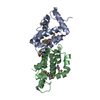
| |||||||||||||||
| Unit cell |
| |||||||||||||||
| Components on special symmetry positions |
| |||||||||||||||
| Details | 2 dimers molecules present in asymmetric unit between monomers C and D; and A and B |
-Components
| #1: Protein | Mass: 16757.527 Da / Num. of mol.: 4 / Fragment: UNP residues 87-229 / Mutation: C205S Source method: isolated from a genetically manipulated source Source: (gene. exp.) Vaccinia virus / Strain: Western Reserve / Gene: VACWR172, A46R / Production host:  #2: Chemical |   Mass: 22.990 Da / Num. of mol.: 2 / Source method: obtained synthetically / Formula: Na Mass: 22.990 Da / Num. of mol.: 2 / Source method: obtained synthetically / Formula: Na#3: Chemical | ChemComp-BR / |   Mass: 79.904 Da / Num. of mol.: 1 / Source method: obtained synthetically / Formula: Br Mass: 79.904 Da / Num. of mol.: 1 / Source method: obtained synthetically / Formula: Br#4: Water | ChemComp-HOH / |  Mass: 18.015 Da / Num. of mol.: 229 / Source method: isolated from a natural source / Formula: H2O Mass: 18.015 Da / Num. of mol.: 229 / Source method: isolated from a natural source / Formula: H2OHas protein modification | Y | |
|---|
-Experimental details
-Experiment
| Experiment | Method: X-RAY DIFFRACTION / Number of used crystals: 1 |
|---|
- Sample preparation
| Crystal | Density Matthews: 2.96 Å3/Da / Density % sol: 58.51 % |
|---|---|
| Crystal grow | Temperature: 295.15 K / Method: vapor diffusion / pH: 7.5 Details: 100 mM bis-tris-propane pH 7.5, 150 mM NaBr, 18% PEG3350, VAPOR DIFFUSION, temperature 295.15K |
-Data collection
| Diffraction | Mean temperature: 100 K |
|---|---|
| Diffraction source | Source: SYNCHROTRON / Site: ESRF  / Beamline: ID29 / Wavelength: 0.9793 Å / Beamline: ID29 / Wavelength: 0.9793 Å |
| Detector | Type: DECTRIS PILATUS 6M / Detector: PIXEL / Date: Feb 14, 2013 |
| Radiation | Monochromator: Si(311) / Protocol: SINGLE WAVELENGTH / Monochromatic (M) / Laue (L): M / Scattering type: x-ray |
| Radiation wavelength | Wavelength: 0.9793 Å / Relative weight: 1 |
| Reflection | Resolution: 1.99→75.924 Å / Num. all: 54679 / Num. obs: 54421 / % possible obs: 99.5 % / Observed criterion σ(F): 0 / Observed criterion σ(I): 0 / Redundancy: 7.9 % / Biso Wilson estimate: 24.9 Å2 / Rmerge(I) obs: 0.163 / Net I/σ(I): 10.4 |
| Reflection shell | Resolution: 1.99→2.04 Å / Redundancy: 7.8 % / Rmerge(I) obs: 1.809 / Mean I/σ(I) obs: 1.3 / Num. unique all: 3762 / % possible all: 92.4 |
- Processing
| Software |
| ||||||||||||||||||||||||||||||||||||||||||||||||||||||||||||||||||||||||||||||||||||||||||||||||||||||||||||||||||||||||||||||||||||||||||||||||||||||||||||||||||||||||||||||||||||||
|---|---|---|---|---|---|---|---|---|---|---|---|---|---|---|---|---|---|---|---|---|---|---|---|---|---|---|---|---|---|---|---|---|---|---|---|---|---|---|---|---|---|---|---|---|---|---|---|---|---|---|---|---|---|---|---|---|---|---|---|---|---|---|---|---|---|---|---|---|---|---|---|---|---|---|---|---|---|---|---|---|---|---|---|---|---|---|---|---|---|---|---|---|---|---|---|---|---|---|---|---|---|---|---|---|---|---|---|---|---|---|---|---|---|---|---|---|---|---|---|---|---|---|---|---|---|---|---|---|---|---|---|---|---|---|---|---|---|---|---|---|---|---|---|---|---|---|---|---|---|---|---|---|---|---|---|---|---|---|---|---|---|---|---|---|---|---|---|---|---|---|---|---|---|---|---|---|---|---|---|---|---|---|---|
| Refinement | Method to determine structure: SAD / Resolution: 1.99→42.46 Å / Cor.coef. Fo:Fc: 0.952 / Cor.coef. Fo:Fc free: 0.942 / SU B: 3.731 / SU ML: 0.101 / Cross valid method: THROUGHOUT / σ(I): 0 / ESU R: 0.151 / ESU R Free: 0.134 / Stereochemistry target values: MAXIMUM LIKELIHOOD / Details: HYDROGENS HAVE BEEN ADDED IN THE RIDING POSITIONS
| ||||||||||||||||||||||||||||||||||||||||||||||||||||||||||||||||||||||||||||||||||||||||||||||||||||||||||||||||||||||||||||||||||||||||||||||||||||||||||||||||||||||||||||||||||||||
| Solvent computation | Ion probe radii: 0.8 Å / Shrinkage radii: 0.8 Å / VDW probe radii: 1.2 Å / Solvent model: MASK | ||||||||||||||||||||||||||||||||||||||||||||||||||||||||||||||||||||||||||||||||||||||||||||||||||||||||||||||||||||||||||||||||||||||||||||||||||||||||||||||||||||||||||||||||||||||
| Displacement parameters | Biso mean: 30.764 Å2
| ||||||||||||||||||||||||||||||||||||||||||||||||||||||||||||||||||||||||||||||||||||||||||||||||||||||||||||||||||||||||||||||||||||||||||||||||||||||||||||||||||||||||||||||||||||||
| Refinement step | Cycle: LAST / Resolution: 1.99→42.46 Å
| ||||||||||||||||||||||||||||||||||||||||||||||||||||||||||||||||||||||||||||||||||||||||||||||||||||||||||||||||||||||||||||||||||||||||||||||||||||||||||||||||||||||||||||||||||||||
| Refine LS restraints |
|