 Movie
Movie Controller

[English] 日本語
 Yorodumi
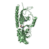
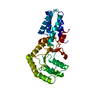
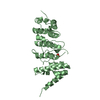
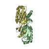
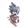
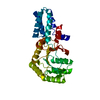
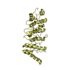
- PDB-6l93: X-ray structure of the ligand-free human TRPV1 ankyrin repeat domain -
+ Open data

- Basic information
| Entry | Database: PDB / ID: 6l93 | ||||||
|---|---|---|---|---|---|---|---|
| Title | X-ray structure of the ligand-free human TRPV1 ankyrin repeat domain | ||||||
 Components Components | Transient receptor potential cation channel subfamily V member 1 | ||||||
 Keywords Keywords | TRANSPORT PROTEIN / Channel domain | ||||||
| Function / homology |  Function and homology information Function and homology informationchemosensory behavior / response to capsazepine / sensory perception of mechanical stimulus / detection of temperature stimulus involved in thermoception / peptide secretion / temperature-gated ion channel activity / detection of chemical stimulus involved in sensory perception of pain / smooth muscle contraction involved in micturition / fever generation / thermoception ...chemosensory behavior / response to capsazepine / sensory perception of mechanical stimulus / detection of temperature stimulus involved in thermoception / peptide secretion / temperature-gated ion channel activity / detection of chemical stimulus involved in sensory perception of pain / smooth muscle contraction involved in micturition / fever generation / thermoception / excitatory extracellular ligand-gated monoatomic ion channel activity / dendritic spine membrane / diet induced thermogenesis / cellular response to alkaloid / TRP channels / intracellularly gated calcium channel activity / cellular response to ATP / detection of temperature stimulus involved in sensory perception of pain / behavioral response to pain / calcium ion import across plasma membrane / voltage-gated calcium channel activity / cellular response to acidic pH / extracellular ligand-gated monoatomic ion channel activity / phosphatidylinositol binding / lipid metabolic process / phosphoprotein binding / GABA-ergic synapse / calcium channel activity / calcium ion transmembrane transport / transmembrane signaling receptor activity / cellular response to heat / sensory perception of taste / protein homotetramerization / calmodulin binding / postsynaptic membrane / cell surface receptor signaling pathway / negative regulation of transcription by RNA polymerase II / ATP binding / membrane / metal ion binding / plasma membrane Similarity search - Function | ||||||
| Biological species |  Homo sapiens (human) Homo sapiens (human) | ||||||
| Method |  X-RAY DIFFRACTION / SYNCHROTRON / MOLECULAR REPLACEMENT / Resolution: 4.47 Å X-RAY DIFFRACTION / SYNCHROTRON / MOLECULAR REPLACEMENT / Resolution: 4.47 Å | ||||||
 Authors Authors | Tanaka, M. / Hayakawa, K. / Unno, M. | ||||||
 Citation Citation | Journal: Acta Crystallogr.,Sect.F / Year: 2020 Title: Structure determination of the human TRPV1 ankyrin-repeat domain under nonreducing conditions. Authors: Tanaka, M. / Hayakawa, K. / Ogawa, N. / Kurokawa, T. / Kitanishi, K. / Ite, K. / Matsui, T. / Mori, Y. / Unno, M. | ||||||
| History |
|
- Structure visualization
| Structure viewer | Molecule: MolmilJmol/JSmol |
|---|
- Downloads & links
-Download
| PDBx/mmCIF format | 6l93.cif.gz | 341.9 KB | Display | PDBx/mmCIF format |
|---|---|---|---|---|
| PDB format | pdb6l93.ent.gz | 241.4 KB | Display | PDB format |
| PDBx/mmJSON format | 6l93.json.gz | Tree view | PDBx/mmJSON format | |
| Others |  Other downloads Other downloads |
-Validation report
| Arichive directory | https://data.pdbj.org/pub/pdb/validation_reports/l9/6l93ftp://data.pdbj.org/pub/pdb/validation_reports/l9/6l93 | HTTPS FTP |
|---|
-Related structure data
| Related structure data | 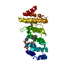 2pnnS S: Starting model for refinement |
|---|---|
| Similar structure data |


-Links
 PDBj
PDBj

- Assembly
| Deposited unit | 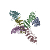
| ||||||||||||
|---|---|---|---|---|---|---|---|---|---|---|---|---|---|
| 1 | 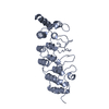
| ||||||||||||
| 2 | 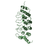
| ||||||||||||
| 3 | 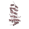
| ||||||||||||
| 4 | 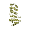
| ||||||||||||
| 5 | 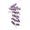
| ||||||||||||
| 6 | 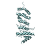
| ||||||||||||
| Unit cell |
|
-Components
| #1: Protein | Mass: 30833.143 Da / Num. of mol.: 6 Source method: isolated from a genetically manipulated source Source: (gene. exp.) Homo sapiens (human) / Gene: TRPV1, VR1 / Production host:  |
|---|
-Experimental details
-Experiment
| Experiment | Method: X-RAY DIFFRACTION / Number of used crystals: 1 |
|---|
- Sample preparation
| Crystal | Density Matthews: 4.46 Å3/Da / Density % sol: 72.39 % |
|---|---|
| Crystal grow | Temperature: 277 K / Method: vapor diffusion, hanging drop / pH: 5 Details: 0.125 M sodium citrate (pH 5.0), 5% PEG 8000, 2.5% (v/v) glycerol |
-Data collection
| Diffraction | Mean temperature: 95 K / Serial crystal experiment: N |
|---|---|
| Diffraction source | Source: SYNCHROTRON / Site: Photon Factory  / Beamline: BL-17A / Wavelength: 0.98 Å / Beamline: BL-17A / Wavelength: 0.98 Å |
| Detector | Type: DECTRIS EIGER X 16M / Detector: PIXEL / Date: Jun 25, 2019 |
| Radiation | Protocol: SINGLE WAVELENGTH / Monochromatic (M) / Laue (L): M / Scattering type: x-ray |
| Radiation wavelength | Wavelength: 0.98 Å / Relative weight: 1 |
| Reflection | Resolution: 4.47→32.95 Å / Num. obs: 19438 / % possible obs: 98.7 % / Redundancy: 3.4 % / Biso Wilson estimate: 179.57 Å2 / CC1/2: 0.993 / Rsym value: 0.098 / Net I/σ(I): 6.4 |
| Reflection shell | Resolution: 4.47→4.63 Å / Num. unique obs: 4678 / CC1/2: 0.817 |
- Processing
| Software |
| ||||||||||||||||||||||||||||||||||||||||||||||||||||||||
|---|---|---|---|---|---|---|---|---|---|---|---|---|---|---|---|---|---|---|---|---|---|---|---|---|---|---|---|---|---|---|---|---|---|---|---|---|---|---|---|---|---|---|---|---|---|---|---|---|---|---|---|---|---|---|---|---|---|
| Refinement | Method to determine structure: MOLECULAR REPLACEMENT Starting model: 2PNN Resolution: 4.47→32.95 Å / SU ML: 0.5466 / Cross valid method: FREE R-VALUE / σ(F): 1.37 / Phase error: 31.6591 Stereochemistry target values: GeoStd + Monomer Library + CDL v1.2
| ||||||||||||||||||||||||||||||||||||||||||||||||||||||||
| Solvent computation | Shrinkage radii: 0.9 Å / VDW probe radii: 1.11 Å / Solvent model: FLAT BULK SOLVENT MODEL | ||||||||||||||||||||||||||||||||||||||||||||||||||||||||
| Displacement parameters | Biso mean: 184.71 Å2 | ||||||||||||||||||||||||||||||||||||||||||||||||||||||||
| Refinement step | Cycle: LAST / Resolution: 4.47→32.95 Å
| ||||||||||||||||||||||||||||||||||||||||||||||||||||||||
| Refine LS restraints |
| ||||||||||||||||||||||||||||||||||||||||||||||||||||||||
| LS refinement shell |
|
