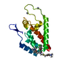
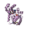
- PDB-3pc7: X-ray crystal structure of the DNA ligase III-alpha BRCT domain. -
+
Open data
ID or keywords:
Loading...
-
Basic information
Entry
Database: PDB / ID: 3pc7
Title
X-ray crystal structure of the DNA ligase III-alpha BRCT domain.
Components
DNA ligase 3
Keywords
LIGASE / DNA repair / BRCT domain / Protein:protein interactions / XRCC1 BRCT2 domain / DNA BINDING PROTEIN
Function / homology
Function and homology information
DNA ligase III-XRCC1 complex / negative regulation of mitochondrial DNA replication / DNA ligase activity / DNA ligase (ATP) / DNA ligase (ATP) activity / Strand-asynchronous mitochondrial DNA replication / double-strand break repair via alternative nonhomologous end joining / lagging strand elongation / HDR through MMEJ (alt-NHEJ) / Resolution of AP sites via the single-nucleotide replacement pathway ...DNA ligase III-XRCC1 complex / negative regulation of mitochondrial DNA replication / DNA ligase activity / DNA ligase (ATP) / DNA ligase (ATP) activity / Strand-asynchronous mitochondrial DNA replication / double-strand break repair via alternative nonhomologous end joining / lagging strand elongation / HDR through MMEJ (alt-NHEJ) / Resolution of AP sites via the single-nucleotide replacement pathway / mitochondrial DNA repair / DNA biosynthetic process / APEX1-Independent Resolution of AP Sites via the Single Nucleotide Replacement Pathway / base-excision repair, gap-filling / Gap-filling DNA repair synthesis and ligation in GG-NER / mitochondrion organization / base-excision repair / double-strand break repair via homologous recombination / Gap-filling DNA repair synthesis and ligation in TC-NER / double-strand break repair / mitochondrial matrix / cell division / mitochondrion / DNA binding / zinc ion binding / nucleoplasm / ATP binding / nucleus Similarity search - Function
DNA ligase 3, BRCT domain / DNA ligase 3 BRCT domain / : / BRCT domain / DNA ligase, ATP-dependent / DNA ligase, ATP-dependent, N-terminal / DNA ligase, ATP-dependent, N-terminal domain superfamily / DNA ligase N terminus / ATP-dependent DNA ligase AMP-binding site. / ATP-dependent DNA ligase signature 2. ...DNA ligase 3, BRCT domain / DNA ligase 3 BRCT domain / : / BRCT domain / DNA ligase, ATP-dependent / DNA ligase, ATP-dependent, N-terminal / DNA ligase, ATP-dependent, N-terminal domain superfamily / DNA ligase N terminus / ATP-dependent DNA ligase AMP-binding site. / ATP-dependent DNA ligase signature 2. / DNA ligase, ATP-dependent, C-terminal / ATP dependent DNA ligase C terminal region / Zinc finger poly(ADP-ribose) polymerase (PARP)-type signature. / Zinc finger, PARP-type superfamily / Poly(ADP-ribose) polymerase and DNA-Ligase Zn-finger region / Zinc finger poly(ADP-ribose) polymerase (PARP)-type profile. / Poly(ADP-ribose) polymerase and DNA-Ligase Zn-finger region / DNA ligase, ATP-dependent, conserved site / Zinc finger, PARP-type / ATP-dependent DNA ligase family profile. / DNA ligase, ATP-dependent, central / ATP dependent DNA ligase domain / breast cancer carboxy-terminal domain / BRCT domain profile. / BRCT domain / BRCT domain superfamily / Nucleic acid-binding, OB-fold / Rossmann fold / 3-Layer(aba) Sandwich / Alpha Beta Similarity search - Domain/homology
In the structure databanks used in Yorodumi, some data are registered as the other names, "COVID-19 virus" and "2019-nCoV". Here are the details of the virus and the list of structure data.
Jan 31, 2019. EMDB accession codes are about to change! (news from PDBe EMDB page)
EMDB accession codes are about to change! (news from PDBe EMDB page)
The allocation of 4 digits for EMDB accession codes will soon come to an end. Whilst these codes will remain in use, new EMDB accession codes will include an additional digit and will expand incrementally as the available range of codes is exhausted. The current 4-digit format prefixed with “EMD-” (i.e. EMD-XXXX) will advance to a 5-digit format (i.e. EMD-XXXXX), and so on. It is currently estimated that the 4-digit codes will be depleted around Spring 2019, at which point the 5-digit format will come into force.
The EM Navigator/Yorodumi systems omit the EMD- prefix.
Related info.:Q: What is EMD? / ID/Accession-code notation in Yorodumi/EM Navigator
Yorodumi is a browser for structure data from EMDB, PDB, SASBDB, etc.
This page is also the successor to EM Navigator detail page, and also detail information page/front-end page for Omokage search.
The word "yorodu" (or yorozu) is an old Japanese word meaning "ten thousand". "mi" (miru) is to see.
Related info.:EMDB / PDB / SASBDB / Comparison of 3 databanks / Yorodumi Search / Aug 31, 2016. New EM Navigator & Yorodumi / Yorodumi Papers / Jmol/JSmol / Function and homology information / Changes in new EM Navigator and Yorodumi
 Movie
Movie Controller


 Open data
 Basic information
 Components
 Keywords
 Function and homology information
 Homo sapiens (human)
 X-RAY DIFFRACTION /
  Authors
 Citation
 Structure visualization
 Downloads & links
 Other downloads


 PDBj
PDBj


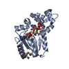
 Assembly


 Mass: 18.015 Da / Num. of mol.: 171 / Source method: isolated from a natural source / Formula: H2O
 Sample preparation
 / Beamline: 22-ID / Wavelength: 0.9793, 0.9748, 0.9795
 Processing