 Movie
Movie Controller


+ Open data

- Basic information
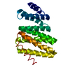
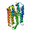
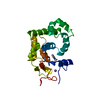
| Entry | Database: PDB / ID: 1gu8 | ||||||
|---|---|---|---|---|---|---|---|
| Title | SENSORY RHODOPSIN II | ||||||
 Components Components | SENSORY RHODOPSIN II | ||||||
 Keywords Keywords | MEMBRANE PROTEIN / ARCHAEAL RHODOPSIN / PHOTORECEPTOR / PHOTOTAXIS | ||||||
| Function / homology |  Function and homology information Function and homology informationphotoreceptor activity / phototransduction / monoatomic ion channel activity / plasma membrane Similarity search - Function | ||||||
| Biological species |  NATRONOMONAS PHARAONIS (archaea) NATRONOMONAS PHARAONIS (archaea) | ||||||
| Method |  X-RAY DIFFRACTION / SYNCHROTRON / MOLECULAR REPLACEMENT / Resolution: 2.27 Å X-RAY DIFFRACTION / SYNCHROTRON / MOLECULAR REPLACEMENT / Resolution: 2.27 Å | ||||||
 Authors Authors | Edman, K. / Royant, A. / Nollert, P. / Maxwell, C.A. / Pebay-Peyroula, E. / Navarro, J. / Neutze, R. / Landau, E.M. | ||||||
 Citation Citation | Journal: Structure / Year: 2002 Title: Early Structural Rearrangements in the Photocycle of an Integral Membrane Sensory Receptor Authors: Edman, K. / Royant, A. / Nollert, P. / Maxwell, C.A. / Pebay-Peyroula, E. / Navarro, J. / Neutze, R. / Landau, E.M. | ||||||
| History |
| ||||||
| Remark 700 | SHEET THE SHEET STRUCTURE OF THIS MOLECULE IS BIFURCATED. IN ORDER TO REPRESENT THIS FEATURE IN ... SHEET THE SHEET STRUCTURE OF THIS MOLECULE IS BIFURCATED. IN ORDER TO REPRESENT THIS FEATURE IN THE SHEET RECORDS BELOW, TWO SHEETS ARE DEFINED. |
- Structure visualization
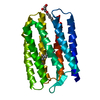
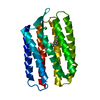
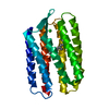
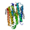
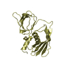
| Structure viewer | Molecule: MolmilJmol/JSmol |
|---|
- Downloads & links
-Download
| PDBx/mmCIF format | 1gu8.cif.gz | 90.4 KB | Display | PDBx/mmCIF format |
|---|---|---|---|---|
| PDB format | pdb1gu8.ent.gz | 70.7 KB | Display | PDB format |
| PDBx/mmJSON format | 1gu8.json.gz | Tree view | PDBx/mmJSON format | |
| Others |  Other downloads Other downloads |
-Validation report
| Arichive directory | https://data.pdbj.org/pub/pdb/validation_reports/gu/1gu8ftp://data.pdbj.org/pub/pdb/validation_reports/gu/1gu8 | HTTPS FTP |
|---|
-Related structure data
| Related structure data |  1gueC  1h68S S: Starting model for refinement C: citing same article ( |
|---|---|
| Similar structure data |


-Links
 PDBj
PDBj


- Assembly
| Deposited unit | 
| ||||||||
|---|---|---|---|---|---|---|---|---|---|
| 1 |
| ||||||||
| Unit cell |
| ||||||||
| Number of models | 2 |
-Components
| #1: Protein | Mass: 25368.814 Da / Num. of mol.: 1 Source method: isolated from a genetically manipulated source Details: MODEL 1 - GROUND STATE, MODEL 2 - K-STATE / Source: (gene. exp.) NATRONOMONAS PHARAONIS (archaea) / Production host:  |
|---|---|
| #2: Chemical | ChemComp-RET /   Mass: 284.436 Da / Num. of mol.: 1 / Source method: obtained synthetically / Formula: C20H28O Mass: 284.436 Da / Num. of mol.: 1 / Source method: obtained synthetically / Formula: C20H28O |
| #3: Chemical | ChemComp-CL /   Mass: 35.453 Da / Num. of mol.: 1 / Source method: obtained synthetically / Formula: Cl Mass: 35.453 Da / Num. of mol.: 1 / Source method: obtained synthetically / Formula: Cl |
| #4: Water | ChemComp-HOH /  Mass: 18.015 Da / Num. of mol.: 16 / Source method: isolated from a natural source / Formula: H2O Mass: 18.015 Da / Num. of mol.: 16 / Source method: isolated from a natural source / Formula: H2O |
-Experimental details
-Experiment
| Experiment | Method: X-RAY DIFFRACTION / Number of used crystals: 1 |
|---|
- Sample preparation
| Crystal | Density Matthews: 2.75 Å3/Da / Density % sol: 53 % Description: SIDE CHAINS, LOOPS AND THE RETINAL WERE OMITTED IN THE STARTING MODEL FOR MOLECULAR REPLACEMENT. LOOPS AB AND CD WERE RESTRAINED IN THE PARTIAL OCCUPANCY REFINEMENT. | ||||||||||||||||||||||||||||||
|---|---|---|---|---|---|---|---|---|---|---|---|---|---|---|---|---|---|---|---|---|---|---|---|---|---|---|---|---|---|---|---|
| Crystal grow | pH: 4.6 / Details: pH 4.6 | ||||||||||||||||||||||||||||||
| Crystal grow | *PLUS Method: unknownDetails: Royant, A., (2001) Proc. Natl. Acad. Sci. U.S.A., 98, 10131. | ||||||||||||||||||||||||||||||
| Components of the solutions | *PLUS
|
-Data collection
| Diffraction | Mean temperature: 100 K |
|---|---|
| Diffraction source | Source: SYNCHROTRON / Site: ESRF  / Beamline: ID14-3 / Wavelength: 0.934 / Beamline: ID14-3 / Wavelength: 0.934 |
| Detector | Type: MARRESEARCH / Detector: CCD / Date: Jun 15, 2001 |
| Radiation | Protocol: SINGLE WAVELENGTH / Monochromatic (M) / Laue (L): M / Scattering type: x-ray |
| Radiation wavelength | Wavelength: 0.934 Å / Relative weight: 1 |
| Reflection | Resolution: 2.27→35.6 Å / Num. obs: 13086 / % possible obs: 99.4 % / Redundancy: 5.8 % / Biso Wilson estimate: 24.1 Å2 / Rmerge(I) obs: 0.139 / Net I/σ(I): 11.5 |
| Reflection shell | Resolution: 2.27→2.38 Å / Redundancy: 5 % / Rmerge(I) obs: 0.764 / Mean I/σ(I) obs: 1.9 / % possible all: 98.1 |
| Reflection | *PLUS Highest resolution: 2.3 Å / Lowest resolution: 36 Å / Num. measured all: 76030 |
| Reflection shell | *PLUS Highest resolution: 2.3 Å / % possible obs: 98.1 % |
- Processing
| Software |
| ||||||||||||||||||||||||||||||||||||||||||||||||||||||||||||||||||||||||||||||||
|---|---|---|---|---|---|---|---|---|---|---|---|---|---|---|---|---|---|---|---|---|---|---|---|---|---|---|---|---|---|---|---|---|---|---|---|---|---|---|---|---|---|---|---|---|---|---|---|---|---|---|---|---|---|---|---|---|---|---|---|---|---|---|---|---|---|---|---|---|---|---|---|---|---|---|---|---|---|---|---|---|---|
| Refinement | Method to determine structure: MOLECULAR REPLACEMENT Starting model: PDB ENTRY 1H68 Resolution: 2.27→36 Å / Rfactor Rfree error: 0.009 / Data cutoff high absF: 10000 / Isotropic thermal model: RESTRAINED / Cross valid method: THROUGHOUT / σ(F): 0
| ||||||||||||||||||||||||||||||||||||||||||||||||||||||||||||||||||||||||||||||||
| Solvent computation | Solvent model: FLAT MODEL / Bsol: 81.5 Å2 / ksol: 0.46 e/Å3 | ||||||||||||||||||||||||||||||||||||||||||||||||||||||||||||||||||||||||||||||||
| Displacement parameters | Biso mean: 29.9 Å2
| ||||||||||||||||||||||||||||||||||||||||||||||||||||||||||||||||||||||||||||||||
| Refine analyze |
| ||||||||||||||||||||||||||||||||||||||||||||||||||||||||||||||||||||||||||||||||
| Refinement step | Cycle: LAST / Resolution: 2.27→36 Å
| ||||||||||||||||||||||||||||||||||||||||||||||||||||||||||||||||||||||||||||||||
| Refine LS restraints |
| ||||||||||||||||||||||||||||||||||||||||||||||||||||||||||||||||||||||||||||||||
| LS refinement shell | Resolution: 2.27→2.41 Å / Rfactor Rfree error: 0.024 / Total num. of bins used: 6
| ||||||||||||||||||||||||||||||||||||||||||||||||||||||||||||||||||||||||||||||||
| Xplor file |
| ||||||||||||||||||||||||||||||||||||||||||||||||||||||||||||||||||||||||||||||||
| Refinement | *PLUS % reflection Rfree: 5.5 % / Rfactor obs: 0.23 / Rfactor Rfree: 0.255 / Rfactor Rwork: 0.23 / Highest resolution: 2.3 Å / Lowest resolution: 36 Å | ||||||||||||||||||||||||||||||||||||||||||||||||||||||||||||||||||||||||||||||||
| Solvent computation | *PLUS | ||||||||||||||||||||||||||||||||||||||||||||||||||||||||||||||||||||||||||||||||
| Displacement parameters | *PLUS | ||||||||||||||||||||||||||||||||||||||||||||||||||||||||||||||||||||||||||||||||
| Refine LS restraints | *PLUS
| ||||||||||||||||||||||||||||||||||||||||||||||||||||||||||||||||||||||||||||||||
| LS refinement shell | *PLUS Rfactor obs: 0.259 |
