 Movie
Movie Controller

[English] 日本語
 Yorodumi
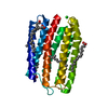
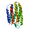
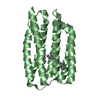
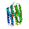
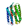
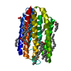
- PDB-1jgj: CRYSTAL STRUCTURE OF SENSORY RHODOPSIN II AT 2.4 ANGSTROMS: INSIG... -
+ Open data

- Basic information
| Entry | Database: PDB / ID: 1jgj | ||||||
|---|---|---|---|---|---|---|---|
| Title | CRYSTAL STRUCTURE OF SENSORY RHODOPSIN II AT 2.4 ANGSTROMS: INSIGHTS INTO COLOR TUNING AND TRANSDUCER INTERACTION | ||||||
 Components Components | SENSORY RHODOPSIN II | ||||||
 Keywords Keywords | SIGNALING PROTEIN / SENSORY RHODOPSIN / MEMBRANE PROTEIN / PHOTOTAXIS RECEPTOR | ||||||
| Function / homology |  Function and homology information Function and homology informationphotoreceptor activity / phototransduction / monoatomic ion channel activity / plasma membrane Similarity search - Function | ||||||
| Biological species |  Natronomonas pharaonis (archaea) Natronomonas pharaonis (archaea) | ||||||
| Method |  X-RAY DIFFRACTION / SYNCHROTRON / MOLECULAR REPLACEMENT / Resolution: 2.4 Å X-RAY DIFFRACTION / SYNCHROTRON / MOLECULAR REPLACEMENT / Resolution: 2.4 Å | ||||||
 Authors Authors | Luecke, H. | ||||||
 Citation Citation | Journal: Science / Year: 2001 Title: Crystal structure of sensory rhodopsin II at 2.4 angstroms: insights into color tuning and transducer interaction. Authors: Luecke, H. / Schobert, B. / Lanyi, J.K. / Spudich, E.N. / Spudich, J.L. #1: Journal: J.Mol.Biol. / Year: 1999Title: Structure of Bacteriorhodopsin at 1.55 Angstrom Resolution Authors: Luecke, H. / Schobert, B. / Richter, H.T. / Cartailler, J.P. / Lanyi, J.K. | ||||||
| History |
|
- Structure visualization
| Structure viewer | Molecule: MolmilJmol/JSmol |
|---|
- Downloads & links
-Download
| PDBx/mmCIF format | 1jgj.cif.gz | 54 KB | Display | PDBx/mmCIF format |
|---|---|---|---|---|
| PDB format | pdb1jgj.ent.gz | 38.5 KB | Display | PDB format |
| PDBx/mmJSON format | 1jgj.json.gz | Tree view | PDBx/mmJSON format | |
| Others |  Other downloads Other downloads |
-Validation report
| Arichive directory | https://data.pdbj.org/pub/pdb/validation_reports/jg/1jgjftp://data.pdbj.org/pub/pdb/validation_reports/jg/1jgj | HTTPS FTP |
|---|
-Related structure data
| Related structure data | 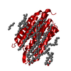 1c3wS S: Starting model for refinement |
|---|---|
| Similar structure data |


-Links
 PDBj
PDBj


- Assembly
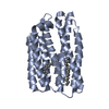
| Deposited unit | 
| ||||||||
|---|---|---|---|---|---|---|---|---|---|
| 1 |
| ||||||||
| Unit cell |
|
-Components
| #1: Protein | Mass: 23175.479 Da / Num. of mol.: 1 Source method: isolated from a genetically manipulated source Source: (gene. exp.) Natronomonas pharaonis (archaea) / Production host: Natronomonas pharaonis (archaea) / References: UniProt: P42196 |
|---|---|
| #2: Sugar | ChemComp-BOG /   Type: D-saccharide / Mass: 292.369 Da / Num. of mol.: 1 Type: D-saccharide / Mass: 292.369 Da / Num. of mol.: 1Source method: isolated from a genetically manipulated source Formula: C14H28O6 / Comment: detergent*YM |
| #3: Chemical | ChemComp-RET /   Mass: 284.436 Da / Num. of mol.: 1 / Source method: obtained synthetically / Formula: C20H28O Mass: 284.436 Da / Num. of mol.: 1 / Source method: obtained synthetically / Formula: C20H28O |
| #4: Water | ChemComp-HOH /  Mass: 18.015 Da / Num. of mol.: 11 / Source method: isolated from a natural source / Formula: H2O Mass: 18.015 Da / Num. of mol.: 11 / Source method: isolated from a natural source / Formula: H2O |
| Has protein modification | Y |
-Experimental details
-Experiment
| Experiment | Method: X-RAY DIFFRACTION / Number of used crystals: 1 |
|---|
- Sample preparation
| Crystal | Density Matthews: 3.13 Å3/Da / Density % sol: 60.75 % / Description: USED PROTEIN WITHOUT RETINAL | |||||||||||||||||||||||||
|---|---|---|---|---|---|---|---|---|---|---|---|---|---|---|---|---|---|---|---|---|---|---|---|---|---|---|
| Crystal grow | Temperature: 295 K / Method: cubic lipid phase / pH: 5.3 Details: CUBIC LIPID PHASE WITH MO, PRECIPITANT KCL, pH 5.3, cubic lipid phase, temperature 295K | |||||||||||||||||||||||||
| Crystal grow | *PLUS Temperature: 20 ℃ / pH: 5.6 Details: Landau, E.M., (1996) Proc.Natl.Acad.Sci.USA., 93, 14532. | |||||||||||||||||||||||||
| Components of the solutions | *PLUS
|
-Data collection
| Diffraction | Mean temperature: 100 K |
|---|---|
| Diffraction source | Source: SYNCHROTRON / Site: ESRF  / Beamline: ID13 / Wavelength: 0.9755 / Beamline: ID13 / Wavelength: 0.9755 |
| Detector | Type: MARRESEARCH / Detector: CCD / Date: Apr 4, 2001 / Details: ID13 |
| Radiation | Monochromator: DOUBLE-XTAL MONO / Protocol: SINGLE WAVELENGTH / Monochromatic (M) / Laue (L): M / Scattering type: x-ray |
| Radiation wavelength | Wavelength: 0.9755 Å / Relative weight: 1 |
| Reflection | Resolution: 2.4→20 Å / Num. obs: 10704 / % possible obs: 92.2 % / Observed criterion σ(I): 0 / Redundancy: 7.5 % / Rsym value: 0.085 / Net I/σ(I): 5.7 |
| Reflection shell | Resolution: 2.4→2.44 Å / Rmerge(I) obs: 0.44 / % possible all: 92 |
| Reflection | *PLUS Highest resolution: 2.4 Å / Lowest resolution: 20 Å / Observed criterion σ(I): 0 / Redundancy: 7.5 % / Num. measured all: 80761 / Rmerge(I) obs: 0.095 |
| Reflection shell | *PLUS % possible obs: 92 % / Num. unique obs: 520 / Rmerge(I) obs: 0.44 |
- Processing
| Software |
| ||||||||||||||||||||||||||||||||||||||||||||||||||||||||||||||||||||||||||||||||
|---|---|---|---|---|---|---|---|---|---|---|---|---|---|---|---|---|---|---|---|---|---|---|---|---|---|---|---|---|---|---|---|---|---|---|---|---|---|---|---|---|---|---|---|---|---|---|---|---|---|---|---|---|---|---|---|---|---|---|---|---|---|---|---|---|---|---|---|---|---|---|---|---|---|---|---|---|---|---|---|---|---|
| Refinement | Method to determine structure: MOLECULAR REPLACEMENT Starting model: 1C3W Resolution: 2.4→20 Å / Cross valid method: THROUGHOUT / σ(F): 0 / Stereochemistry target values: ENGH & HUBER, CSD
| ||||||||||||||||||||||||||||||||||||||||||||||||||||||||||||||||||||||||||||||||
| Displacement parameters |
| ||||||||||||||||||||||||||||||||||||||||||||||||||||||||||||||||||||||||||||||||
| Refinement step | Cycle: LAST / Resolution: 2.4→20 Å
| ||||||||||||||||||||||||||||||||||||||||||||||||||||||||||||||||||||||||||||||||
| Refine LS restraints |
| ||||||||||||||||||||||||||||||||||||||||||||||||||||||||||||||||||||||||||||||||
| LS refinement shell | Resolution: 2.4→2.45 Å
| ||||||||||||||||||||||||||||||||||||||||||||||||||||||||||||||||||||||||||||||||
| Xplor file | Serial no: 1 / Param file: PROTEIN_REP.PARAM / Topol file: PROTEIN.TOP | ||||||||||||||||||||||||||||||||||||||||||||||||||||||||||||||||||||||||||||||||
| Software | *PLUS Name: CNS / Version: 1 / Classification: refinement | ||||||||||||||||||||||||||||||||||||||||||||||||||||||||||||||||||||||||||||||||
| Refinement | *PLUS Rfactor Rfree: 0.28 | ||||||||||||||||||||||||||||||||||||||||||||||||||||||||||||||||||||||||||||||||
| Solvent computation | *PLUS | ||||||||||||||||||||||||||||||||||||||||||||||||||||||||||||||||||||||||||||||||
| Displacement parameters | *PLUS | ||||||||||||||||||||||||||||||||||||||||||||||||||||||||||||||||||||||||||||||||
| Refine LS restraints | *PLUS
| ||||||||||||||||||||||||||||||||||||||||||||||||||||||||||||||||||||||||||||||||
| LS refinement shell | *PLUS Rfactor Rfree: 0.302 / Rfactor Rwork: 0.29 |
