| Entry | Database: PDB / ID: 5d3d
|
|---|
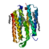
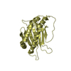
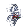
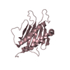
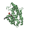
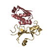
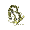
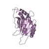
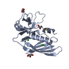
| Title | Crystal structure of Staphylococcal Superantigen-Like protein 3 |
|---|
 Components Components | Staphylococcal Superantigen-Like protein 3 |
|---|
 Keywords Keywords | IMMUNE SYSTEM / Superantigens / Superantigen-like proteins / SSL / SSL3 / Toll-Like Receptor 2 / TLR2 / Immunology / Inflammation / Inhibition |
|---|
| Function / homology |  Function and homology information Function and homology information
Staphylococcus aureus exotoxin / Staphylococcal superantigen-like OB-fold domain / Staphylococcal superantigen-like OB-fold domain / Ubiquitin-like (UB roll) - #120 / Staphylococcal enterotoxin/Streptococcal pyrogenic exotoxin, conserved site / Staphyloccocal enterotoxin/Streptococcal pyrogenic exotoxin signature 2. / Superantigen, staphylococcal/streptococcal toxin, bacterial / Staphylococcal/Streptococcal toxin, beta-grasp domain / Staphylococcal/Streptococcal toxin, beta-grasp domain / Superantigen toxin, C-terminal ...Staphylococcus aureus exotoxin / Staphylococcal superantigen-like OB-fold domain / Staphylococcal superantigen-like OB-fold domain / Ubiquitin-like (UB roll) - #120 / Staphylococcal enterotoxin/Streptococcal pyrogenic exotoxin, conserved site / Staphyloccocal enterotoxin/Streptococcal pyrogenic exotoxin signature 2. / Superantigen, staphylococcal/streptococcal toxin, bacterial / Staphylococcal/Streptococcal toxin, beta-grasp domain / Staphylococcal/Streptococcal toxin, beta-grasp domain / Superantigen toxin, C-terminal / OB fold (Dihydrolipoamide Acetyltransferase, E2P) - #110 / Enterotoxin / Ubiquitin-like (UB roll) / OB fold (Dihydrolipoamide Acetyltransferase, E2P) / Roll / Beta Barrel / Mainly Beta / Alpha BetaSimilarity search - Domain/homology |
|---|
| Biological species |   Staphylococcus aureus (bacteria) Staphylococcus aureus (bacteria) |
|---|
| Method |  X-RAY DIFFRACTION / SYNCHROTRON / MOLECULAR REPLACEMENT / Resolution: 1.94 Å X-RAY DIFFRACTION / SYNCHROTRON / MOLECULAR REPLACEMENT / Resolution: 1.94 Å |
|---|
 Authors Authors | Feitsma, L.J. / Huizinga, E.G. |
|---|
| Funding support |  Netherlands, 3items Netherlands, 3items | Organization | Grant number | Country |
|---|
| Netherlands Organization for Scientific Research | ECHO Grant 700.58.006 | Netherlands | | Netherlands Organization for Health Research and Development | ZonMw Grant 205200004 | Netherlands | | Dutch Top Institute Pharma Project | D1-101 | Netherlands |
|
|---|
 Citation Citation | Journal: Proc.Natl.Acad.Sci.USA / Year: 2015
Title: Structural basis for inhibition of TLR2 by staphylococcal superantigen-like protein 3 (SSL3).
Authors: Koymans, K.J. / Feitsma, L.J. / Brondijk, T.H. / Aerts, P.C. / Lukkien, E. / Lossl, P. / van Kessel, K.P. / de Haas, C.J. / van Strijp, J.A. / Huizinga, E.G. |
|---|
| History | | Deposition | Aug 6, 2015 | Deposition site: RCSB / Processing site: PDBE |
|---|
| Revision 1.0 | Aug 19, 2015 | Provider: repository / Type: Initial release |
|---|
| Revision 1.1 | Sep 2, 2015 | Group: Database references |
|---|
| Revision 1.2 | Sep 9, 2015 | Group: Database references |
|---|
| Revision 1.3 | May 1, 2024 | Group: Data collection / Database references / Refinement description
Category: chem_comp_atom / chem_comp_bond ...chem_comp_atom / chem_comp_bond / database_2 / pdbx_initial_refinement_model
Item: _database_2.pdbx_DOI / _database_2.pdbx_database_accession |
|---|
|
|---|
 Movie
Movie Controller


 Open data
 Basic information
 Structure visualization
 Downloads & links
 Other downloads


 PDBj
PDBj

 Assembly


 Mass: 35.453 Da / Num. of mol.: 4 / Source method: obtained synthetically / Formula: Cl
 Mass: 58.082 Da / Num. of mol.: 2 / Source method: obtained synthetically / Formula: CNS
 Mass: 92.094 Da / Num. of mol.: 5 / Source method: obtained synthetically / Formula: C3H8O3
 Mass: 22.990 Da / Num. of mol.: 1 / Source method: obtained synthetically / Formula: Na
 Sample preparation
 / Beamline: X06SA / Wavelength: 0.9999 Å
 Processing