 Movie
Movie Controller

[English] 日本語
 Yorodumi
- PDB-4j32: Structure of the effector - immunity system Tae4 / Tai4 from Salm... -
+ Open data

- Basic information
| Entry | Database: PDB / ID: 4j32 | ||||||
|---|---|---|---|---|---|---|---|
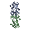
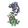
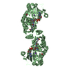
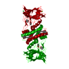
| Title | Structure of the effector - immunity system Tae4 / Tai4 from Salmonella typhimurium | ||||||
 Components Components |
| ||||||
 Keywords Keywords | TOXIN/INHIBITOR / N1pC/P60 papain like cysteine peptidase Tae4 / peptidoglycan hydrolase / immunity protein Tai4 / Tae4: cytoplasmatic / Tai4: periplasmatic / TOXIN-INHIBITOR complex | ||||||
| Function / homology |  Function and homology information Function and homology informationendopeptidase fold (from Nostoc punctiforme) - #70 / Type VI secretion system (T6SS), amidase immunity protein / T6SS superfamily / Type VI secretion system (T6SS), amidase immunity protein / Four Helix Bundle (Hemerythrin (Met), subunit A) - #1620 / Type VI secretion system (T6SS), amidase effector protein 4 / Type VI secretion system (T6SS), amidase effector protein 4 / endopeptidase fold (from Nostoc punctiforme) / Four Helix Bundle (Hemerythrin (Met), subunit A) / Alpha-Beta Complex ...endopeptidase fold (from Nostoc punctiforme) - #70 / Type VI secretion system (T6SS), amidase immunity protein / T6SS superfamily / Type VI secretion system (T6SS), amidase immunity protein / Four Helix Bundle (Hemerythrin (Met), subunit A) - #1620 / Type VI secretion system (T6SS), amidase effector protein 4 / Type VI secretion system (T6SS), amidase effector protein 4 / endopeptidase fold (from Nostoc punctiforme) / Four Helix Bundle (Hemerythrin (Met), subunit A) / Alpha-Beta Complex / Up-down Bundle / Mainly Alpha / Alpha Beta Similarity search - Domain/homology | ||||||
| Biological species |  Salmonella enterica subsp. enterica serovar Typhimurium (bacteria) Salmonella enterica subsp. enterica serovar Typhimurium (bacteria) | ||||||
| Method |  X-RAY DIFFRACTION / SYNCHROTRON / rigid-body refinement / Resolution: 1.8 Å X-RAY DIFFRACTION / SYNCHROTRON / rigid-body refinement / Resolution: 1.8 Å | ||||||
 Authors Authors | Benz, J. / Reinstein, J. / Meinhart, A. | ||||||
 Citation Citation | Journal: Plos One / Year: 2013 Title: Structural Insights into the Effector - Immunity System Tae4/Tai4 from Salmonella typhimurium. Authors: Benz, J. / Reinstein, J. / Meinhart, A. | ||||||
| History |
|
- Structure visualization
| Structure viewer | Molecule: MolmilJmol/JSmol |
|---|
- Downloads & links
-Download
| PDBx/mmCIF format | 4j32.cif.gz | 124.2 KB | Display | PDBx/mmCIF format |
|---|---|---|---|---|
| PDB format | pdb4j32.ent.gz | 98.1 KB | Display | PDB format |
| PDBx/mmJSON format | 4j32.json.gz | Tree view | PDBx/mmJSON format | |
| Others |  Other downloads Other downloads |
-Validation report
| Arichive directory | https://data.pdbj.org/pub/pdb/validation_reports/j3/4j32ftp://data.pdbj.org/pub/pdb/validation_reports/j3/4j32 | HTTPS FTP |
|---|
-Related structure data
| Related structure data |  4j30SC S: Starting model for refinement C: citing same article ( |
|---|---|
| Similar structure data |


-Links
 PDBj
PDBj


- Assembly
| Deposited unit | 
| ||||||||
|---|---|---|---|---|---|---|---|---|---|
| 1 | 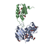
| ||||||||
| Unit cell |
|
-Components
| #1: Protein | Mass: 19369.020 Da / Num. of mol.: 1 Source method: isolated from a genetically manipulated source Source: (gene. exp.) Salmonella enterica subsp. enterica serovar Typhimurium (bacteria)Strain: LT2 / SGSC1412 / ATCC 700720 / Plasmid: pET28b / Production host: | ||||||
|---|---|---|---|---|---|---|---|
| #2: Protein | Mass: 11711.182 Da / Num. of mol.: 1 Fragment: periplasmatic inhibitor domain (UNP residues 27-127) Source method: isolated from a genetically manipulated source Source: (gene. exp.) Salmonella enterica subsp. enterica serovar Typhimurium (bacteria)Strain: LT2 / SGSC1412 / ATCC 700720 / Plasmid: pET28b / Production host: | ||||||
| #3: Chemical |   Mass: 189.100 Da / Num. of mol.: 2 / Source method: obtained synthetically / Formula: C6H5O7 Mass: 189.100 Da / Num. of mol.: 2 / Source method: obtained synthetically / Formula: C6H5O7#4: Chemical | ChemComp-ETX /   Mass: 90.121 Da / Num. of mol.: 8 / Source method: obtained synthetically / Formula: C4H10O2 Mass: 90.121 Da / Num. of mol.: 8 / Source method: obtained synthetically / Formula: C4H10O2#5: Water | ChemComp-HOH / |  Mass: 18.015 Da / Num. of mol.: 202 / Source method: isolated from a natural source / Formula: H2O Mass: 18.015 Da / Num. of mol.: 202 / Source method: isolated from a natural source / Formula: H2OHas protein modification | Y | |
-Experimental details
-Experiment
| Experiment | Method: X-RAY DIFFRACTION / Number of used crystals: 1 |
|---|
- Sample preparation
| Crystal | Density Matthews: 3.44 Å3/Da / Density % sol: 64.24 % |
|---|---|
| Crystal grow | Temperature: 293 K / Method: vapor diffusion, hanging drop / pH: 6 Details: 200 mM tri-sodium citrate pH 6.0, 30 % (v/v) 2-ethoxyethanol , VAPOR DIFFUSION, HANGING DROP, temperature 293K |
-Data collection
| Diffraction | Mean temperature: 100 K |
|---|---|
| Diffraction source | Source: SYNCHROTRON / Site: SLS  / Beamline: X10SA / Wavelength: 0.99981 Å / Beamline: X10SA / Wavelength: 0.99981 Å |
| Detector | Type: PSI PILATUS 6M / Detector: PIXEL / Date: Aug 5, 2012 / Details: Dynamically bendable mirror |
| Radiation | Monochromator: LN2 cooled fixed-exit Si(111) monochromator / Protocol: SINGLE WAVELENGTH / Monochromatic (M) / Laue (L): M / Scattering type: x-ray |
| Radiation wavelength | Wavelength: 0.99981 Å / Relative weight: 1 |
| Reflection | Resolution: 1.8→40 Å / Num. all: 42126 / Num. obs: 42126 / % possible obs: 99.1 % / Observed criterion σ(F): 0 / Observed criterion σ(I): 0 / Redundancy: 18.1 % / Rmerge(I) obs: 0.102 / Net I/σ(I): 18.7 |
| Reflection shell | Resolution: 1.8→1.9 Å / Redundancy: 19 % / Rmerge(I) obs: 0.661 / Mean I/σ(I) obs: 4.7 / Num. unique all: 6089 / % possible all: 98.6 |
- Processing
| Software |
| ||||||||||||||||||||||||||||||||||||||||||||||||||||||||||||||||||||||||||||||||||||||||||
|---|---|---|---|---|---|---|---|---|---|---|---|---|---|---|---|---|---|---|---|---|---|---|---|---|---|---|---|---|---|---|---|---|---|---|---|---|---|---|---|---|---|---|---|---|---|---|---|---|---|---|---|---|---|---|---|---|---|---|---|---|---|---|---|---|---|---|---|---|---|---|---|---|---|---|---|---|---|---|---|---|---|---|---|---|---|---|---|---|---|---|---|
| Refinement | Method to determine structure: rigid-body refinement Starting model: 4J30 Resolution: 1.8→37.9 Å / Cor.coef. Fo:Fc: 0.958 / Cor.coef. Fo:Fc free: 0.947 / SU B: 3.393 / SU ML: 0.056 / Cross valid method: THROUGHOUT / σ(F): 0 / ESU R: 0.094 / ESU R Free: 0.091 / Stereochemistry target values: MAXIMUM LIKELIHOOD / Details: HYDROGENS HAVE BEEN ADDED IN THE RIDING POSITIONS
| ||||||||||||||||||||||||||||||||||||||||||||||||||||||||||||||||||||||||||||||||||||||||||
| Solvent computation | Ion probe radii: 0.8 Å / Shrinkage radii: 0.8 Å / VDW probe radii: 1.2 Å / Solvent model: MASK | ||||||||||||||||||||||||||||||||||||||||||||||||||||||||||||||||||||||||||||||||||||||||||
| Displacement parameters | Biso mean: 26.896 Å2
| ||||||||||||||||||||||||||||||||||||||||||||||||||||||||||||||||||||||||||||||||||||||||||
| Refinement step | Cycle: LAST / Resolution: 1.8→37.9 Å
| ||||||||||||||||||||||||||||||||||||||||||||||||||||||||||||||||||||||||||||||||||||||||||
| Refine LS restraints |
| ||||||||||||||||||||||||||||||||||||||||||||||||||||||||||||||||||||||||||||||||||||||||||
| LS refinement shell | Resolution: 1.8→1.847 Å / Total num. of bins used: 20
| ||||||||||||||||||||||||||||||||||||||||||||||||||||||||||||||||||||||||||||||||||||||||||
| Refinement TLS params. | Method: refined / Refine-ID: X-RAY DIFFRACTION
| ||||||||||||||||||||||||||||||||||||||||||||||||||||||||||||||||||||||||||||||||||||||||||
| Refinement TLS group |
|
