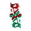
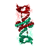
- PDB-3th1: Crystal structure of chlorocatechol 1,2-dioxygenase from Pseudomo... -
+
Open data
ID or keywords:
Loading...
-
Basic information
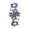
Entry
Database: PDB / ID: 3th1
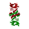
Title
Crystal structure of chlorocatechol 1,2-dioxygenase from Pseudomonas putida
Components
Chlorocatechol 1,2-dioxygenase
Keywords
OXIDOREDUCTASE / Catechol 1 / 2-dioxygenase family / iron binding
Function / homology
Function and homology information
catechol 1,2-dioxygenase activity / Oxidoreductases; Acting on single donors with incorporation of molecular oxygen (oxygenases); With incorporation of two atoms of oxygen / catechol-containing compound metabolic process / ferric iron binding Similarity search - Function
Mass: 29024.676 Da / Num. of mol.: 3 Source method: isolated from a genetically manipulated source Source: (gene. exp.) Pseudomonas putida (bacteria) / Gene: clcA / Plasmid: PTYB2 / Production host: Escherichia coli (E. coli) / Strain (production host): BL21(DE3) References: UniProt: P11451, Oxidoreductases; Acting on single donors with incorporation of molecular oxygen (oxygenases); With incorporation of two atoms of oxygen
Mass: 18.015 Da / Num. of mol.: 17 / Source method: isolated from a natural source / Formula: H2O
-
Experimental details
-
Experiment
Experiment
Method: X-RAY DIFFRACTION / Number of used crystals: 1
-
Sample preparation
Crystal
Density Matthews: 3.34 Å3/Da / Density % sol: 63.2 %
Crystal grow
Temperature: 294 K / Method: vapor diffusion, sitting drop Details: 14% PEG 8000 and 0.2 M magnesium acetate tetrahydrated, VAPOR DIFFUSION, SITTING DROP, temperature 294K
In the structure databanks used in Yorodumi, some data are registered as the other names, "COVID-19 virus" and "2019-nCoV". Here are the details of the virus and the list of structure data.
Jan 31, 2019. EMDB accession codes are about to change! (news from PDBe EMDB page)
EMDB accession codes are about to change! (news from PDBe EMDB page)
The allocation of 4 digits for EMDB accession codes will soon come to an end. Whilst these codes will remain in use, new EMDB accession codes will include an additional digit and will expand incrementally as the available range of codes is exhausted. The current 4-digit format prefixed with “EMD-” (i.e. EMD-XXXX) will advance to a 5-digit format (i.e. EMD-XXXXX), and so on. It is currently estimated that the 4-digit codes will be depleted around Spring 2019, at which point the 5-digit format will come into force.
The EM Navigator/Yorodumi systems omit the EMD- prefix.
Related info.:Q: What is EMD? / ID/Accession-code notation in Yorodumi/EM Navigator
Yorodumi is a browser for structure data from EMDB, PDB, SASBDB, etc.
This page is also the successor to EM Navigator detail page, and also detail information page/front-end page for Omokage search.
The word "yorodu" (or yorozu) is an old Japanese word meaning "ten thousand". "mi" (miru) is to see.
Related info.:EMDB / PDB / SASBDB / Comparison of 3 databanks / Yorodumi Search / Aug 31, 2016. New EM Navigator & Yorodumi / Yorodumi Papers / Jmol/JSmol / Function and homology information / Changes in new EM Navigator and Yorodumi
 Movie
Movie Controller

 Yorodumi
 Open data
 Basic information
 Components
 Keywords
 Function and homology information
 Pseudomonas putida (bacteria)
 X-RAY DIFFRACTION /
  Authors
 Citation
 Structure visualization
 Downloads & links
 Other downloads


 PDBj
PDBj Assembly
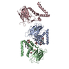


 Mass: 55.845 Da / Num. of mol.: 3 / Source method: obtained synthetically / Formula: Fe
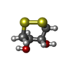
 Mass: 152.235 Da / Num. of mol.: 2 / Source method: obtained synthetically / Formula: C4H8O2S2
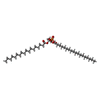
 Mass: 704.998 Da / Num. of mol.: 2 / Source method: obtained synthetically / Formula: C39H77O8P
 Mass: 24.305 Da / Num. of mol.: 2 / Source method: obtained synthetically / Formula: Mg
 Mass: 59.044 Da / Num. of mol.: 1 / Source method: obtained synthetically / Formula: C2H3O2
 Mass: 92.094 Da / Num. of mol.: 2 / Source method: obtained synthetically / Formula: C3H8O3
 Sample preparation
 / Beamline: W01B-MX2 / Wavelength: 1.459 Å
 Processing