[English] 日本語
Yorodumi
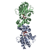
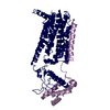
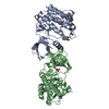
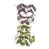
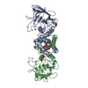
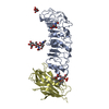
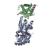
- PDB-6wgz: Crystal structure of HyBcl-2-4 with HyBak1 BH3 -

+
Open data


ID or keywords:

Loading...

-
Basic information

Entry
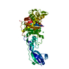
Database: PDB / ID: 6wgz
TitleCrystal structure of HyBcl-2-4 with HyBak1 BH3
Components
  • Bak
  • Bcl-2-like 4
  • Maltodextrin-binding protein
KeywordsAPOPTOSIS / Antiapoptotic / complex with BH3 motif
Function / homology
Function and homology information


carbohydrate transmembrane transporter activity / regulation of apoptotic process / membrane => GO:0016020 / periplasmic space / apoptotic process
Similarity search - Function
Apoptosis regulator, Bcl-2, BH2 motif, conserved site / Apoptosis regulator, Bcl-2 family BH2 motif signature. / BCL (B-Cell lymphoma); contains BH1, BH2 regions / Bcl-2 family / Bcl-2, Bcl-2 homology region 1-3 / Bcl2-like / Apoptosis regulator proteins, Bcl-2 family / BCL2-like apoptosis inhibitors family profile. / Bcl-2-like superfamily / Maltose/Cyclodextrin ABC transporter, substrate-binding protein ...Apoptosis regulator, Bcl-2, BH2 motif, conserved site / Apoptosis regulator, Bcl-2 family BH2 motif signature. / BCL (B-Cell lymphoma); contains BH1, BH2 regions / Bcl-2 family / Bcl-2, Bcl-2 homology region 1-3 / Bcl2-like / Apoptosis regulator proteins, Bcl-2 family / BCL2-like apoptosis inhibitors family profile. / Bcl-2-like superfamily / Maltose/Cyclodextrin ABC transporter, substrate-binding protein / Solute-binding family 1, conserved site / Bacterial extracellular solute-binding proteins, family 1 signature. / Bacterial extracellular solute-binding protein / Bacterial extracellular solute-binding protein
Similarity search - Domain/homology
alpha-maltotriose / ACETATE ION / Maltodextrin-binding protein / Bak / Bcl-2-like 4
Similarity search - Component
Biological speciesEscherichia coli (E. coli)
Hydra vulgaris (swiftwater hydra)
MethodX-RAY DIFFRACTION / SYNCHROTRON / MOLECULAR REPLACEMENT / Resolution: 2.2 Å
AuthorsKvansakul, M. / Hinds, M.G. / Banjara, S.
Funding support Australia, 1items
OrganizationGrant numberCountry
Australian Research Council (ARC)DP190103591 Australia
CitationJournal: To Be Published
Title: Crystal structures of HyBcl-2-4 with HyBak1 BH3 and HyBax BH3
Authors: Banjara, S. / Sa, J.D. / Hinds, M.G. / Kvansakul, M.
History
DepositionApr 7, 2020Deposition site: RCSB / Processing site: RCSB
Revision 1.0Apr 21, 2021Provider: repository / Type: Initial release
Revision 1.1Oct 18, 2023Group: Data collection / Database references / Refinement description
Category: chem_comp_atom / chem_comp_bond ...chem_comp_atom / chem_comp_bond / database_2 / pdbx_initial_refinement_model
Item: _database_2.pdbx_DOI / _database_2.pdbx_database_accession

-
Structure visualization

Structure viewerMolecule:
MolmilJmol/JSmol

Downloads & links

-
Assembly

Deposited unit
A: Maltodextrin-binding protein
B: Bcl-2-like 4
C: Bak
hetero molecules


Theoretical massNumber of molelcules
Total (without water)63,0536
Polymers62,4303
Non-polymers6233
Water2,306128
1
B: Bcl-2-like 4
C: Bak
hetero molecules

A: Maltodextrin-binding protein
hetero molecules


Theoretical massNumber of molelcules
Total (without water)63,0536
Polymers62,4303
Non-polymers6233
Water543
TypeNameSymmetry operationNumber
identity operation1_555x,y,z1
crystal symmetry operation1_455x-1,y,z1
Buried area3890 Å2
ΔGint-24 kcal/mol
Surface area22770 Å2
MethodPISA
Unit cell
Length a, b, c (Å)54.245, 73.650, 140.800
Angle α, β, γ (deg.)90.000, 90.000, 90.000
Int Tables number19
Space group name H-MP212121
Space group name HallP2ac2ab
Symmetry operation#1: x,y,z
#2: x+1/2,-y+1/2,-z
#3: -x,y+1/2,-z+1/2
#4: -x+1/2,-y,z+1/2

-
Components

-
Protein , 2 types, 2 molecules AB

#1: Protein Maltodextrin-binding protein


Mass: 40759.051 Da / Num. of mol.: 1
Source method: isolated from a genetically manipulated source
Source: (gene. exp.) Escherichia coli (E. coli) / Gene: DAH37_23060 / Production host: Escherichia coli (E. coli) / References: UniProt: A0A4Z0THX4
#2: Protein Bcl-2-like 4 / Bcl-2-like protein 1


Mass: 18756.225 Da / Num. of mol.: 1
Source method: isolated from a genetically manipulated source
Source: (gene. exp.) Hydra vulgaris (swiftwater hydra) / Gene: BCL2L1 / Production host: Escherichia coli (E. coli) / References: UniProt: A7LM80

-
Protein/peptide / Sugars , 2 types, 2 molecules C

#3: Protein/peptide Bak


Mass: 2915.112 Da / Num. of mol.: 1 / Source method: obtained synthetically / Source: (synth.) Hydra vulgaris (swiftwater hydra) / References: UniProt: A1E3K5
#4: Polysaccharide alpha-D-glucopyranose-(1-4)-alpha-D-glucopyranose-(1-4)-alpha-D-glucopyranose / alpha-maltotriose


Type: oligosaccharide, Oligosaccharide / Class: Nutrient / Mass: 504.438 Da / Num. of mol.: 1
Source method: isolated from a genetically manipulated source
Details: oligosaccharide / References: alpha-maltotriose
DescriptorTypeProgram
DGlcpa1-4DGlcpa1-4DGlcpa1-ROHGlycam Condensed SequenceGMML 1.0
WURCS=2.0/1,3,2/[a2122h-1a_1-5]/1-1-1/a4-b1_b4-c1WURCSPDB2Glycan 1.1.0
[][a-D-Glcp]{[(4+1)][a-D-Glcp]{[(4+1)][a-D-Glcp]{}}}LINUCSPDB-CARE

-
Non-polymers , 2 types, 130 molecules

#5: Chemical ChemComp-ACT / ACETATE ION


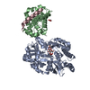
Mass: 59.044 Da / Num. of mol.: 2 / Source method: obtained synthetically / Formula: C2H3O2
#6: Water ChemComp-HOH / water


Mass: 18.015 Da / Num. of mol.: 128 / Source method: isolated from a natural source / Formula: H2O

-
Details

Has ligand of interestN

-
Experimental details

-
Experiment

ExperimentMethod: X-RAY DIFFRACTION / Number of used crystals: 1

-
Sample preparation

CrystalDensity Matthews: 2.25 Å3/Da / Density % sol: 45.4 % / Description: plate
Crystal growTemperature: 293 K / Method: vapor diffusion, sitting drop / pH: 6.8 / Details: 0.2M Magnesium acetate tetra hydrate, 20% PEG 3350

-
Data collection

DiffractionMean temperature: 100 K / Serial crystal experiment: N
Diffraction sourceSource: SYNCHROTRON / Site: Australian Synchrotron / Beamline: MX2 / Wavelength: 0.9537 Å
DetectorType: DECTRIS EIGER X 16M / Detector: PIXEL / Date: Mar 14, 2020
RadiationProtocol: SINGLE WAVELENGTH / Monochromatic (M) / Laue (L): M / Scattering type: x-ray
Radiation wavelengthWavelength: 0.9537 Å / Relative weight: 1
ReflectionResolution: 2.19→43.68 Å / Num. obs: 29115 / % possible obs: 98.49 % / Redundancy: 10.8 % / Biso Wilson estimate: 43.33 Å2 / CC1/2: 0.99 / Rmerge(I) obs: 0.137 / Net I/σ(I): 8.6
Reflection shellResolution: 2.19→2.27 Å / Rmerge(I) obs: 1.56 / Mean I/σ(I) obs: 1.4 / Num. unique obs: 2769 / CC1/2: 0.94

-
Processing

Software
NameVersionClassification
xia2data processing
PHASERphasing
DIALSdata reduction
Aimlessdata scaling
Cootmodel building
PHENIX1.17.1_3660refinement
RefinementMethod to determine structure: MOLECULAR REPLACEMENT
Starting model: 1PQ1
Resolution: 2.2→43.68 Å / SU ML: 0.2578 / Cross valid method: FREE R-VALUE / σ(F): 1.34 / Phase error: 29.9134
RfactorNum. reflection% reflection
Rfree0.2511 1439 4.96 %
Rwork0.2223 --
obs0.2238 29009 98.26 %
Solvent computationShrinkage radii: 0.9 Å / VDW probe radii: 1.11 Å
Displacement parametersBiso mean: 52.84 Å2
Refinement stepCycle: LAST / Resolution: 2.2→43.68 Å
ProteinNucleic acidLigandSolventTotal
Num. atoms4198 0 42 128 4368
Refine LS restraints
Refine-IDTypeDev idealNumber
X-RAY DIFFRACTIONf_bond_d0.00294355
X-RAY DIFFRACTIONf_angle_d0.62425911
X-RAY DIFFRACTIONf_chiral_restr0.038649
X-RAY DIFFRACTIONf_plane_restr0.0027762
X-RAY DIFFRACTIONf_dihedral_angle_d19.96461560
LS refinement shell
Resolution (Å)Rfactor RfreeNum. reflection RfreeRfactor RworkNum. reflection RworkRefine-ID% reflection obs (%)
2.2-2.280.36431300.36582509X-RAY DIFFRACTION90.84
2.28-2.370.35181430.30062732X-RAY DIFFRACTION98.43
2.37-2.470.28071410.27772727X-RAY DIFFRACTION98.86
2.47-2.610.29121470.26462727X-RAY DIFFRACTION99
2.61-2.770.28511490.27512699X-RAY DIFFRACTION98.27
2.77-2.980.30221460.2632778X-RAY DIFFRACTION99.49
2.98-3.280.27571650.25662758X-RAY DIFFRACTION99.15
3.28-3.760.2431280.21092801X-RAY DIFFRACTION99.25
3.76-4.730.19631370.17582856X-RAY DIFFRACTION99.57
4.73-43.680.22491530.19112983X-RAY DIFFRACTION99.52

+
About Yorodumi

-
News

-
Feb 9, 2022. New format data for meta-information of EMDB entries

New format data for meta-information of EMDB entries

  • Version 3 of the EMDB header file is now the official format.
  • The previous official version 1.9 will be removed from the archive.

Related info.:EMDB header

External links:wwPDB to switch to version 3 of the EMDB data model

-
Aug 12, 2020. Covid-19 info

Covid-19 info

URL: https://pdbjlvh1.pdbj.org/emnavi/covid19.php

New page: Covid-19 featured information page in EM Navigator.

Related info.:Covid-19 info / Mar 5, 2020. Novel coronavirus structure data

+
Mar 5, 2020. Novel coronavirus structure data

Novel coronavirus structure data

Related info.:Yorodumi Speices / Aug 12, 2020. Covid-19 info

External links:COVID-19 featured content - PDBj / Molecule of the Month (242):Coronavirus Proteases

+
Jan 31, 2019. EMDB accession codes are about to change! (news from PDBe EMDB page)

EMDB accession codes are about to change! (news from PDBe EMDB page)

  • The allocation of 4 digits for EMDB accession codes will soon come to an end. Whilst these codes will remain in use, new EMDB accession codes will include an additional digit and will expand incrementally as the available range of codes is exhausted. The current 4-digit format prefixed with “EMD-” (i.e. EMD-XXXX) will advance to a 5-digit format (i.e. EMD-XXXXX), and so on. It is currently estimated that the 4-digit codes will be depleted around Spring 2019, at which point the 5-digit format will come into force.
  • The EM Navigator/Yorodumi systems omit the EMD- prefix.

Related info.:Q: What is EMD? / ID/Accession-code notation in Yorodumi/EM Navigator

External links:EMDB Accession Codes are Changing Soon! / Contact to PDBj

+
Jul 12, 2017. Major update of PDB

Major update of PDB

  • wwPDB released updated PDB data conforming to the new PDBx/mmCIF dictionary.
  • This is a major update changing the version number from 4 to 5, and with Remediation, in which all the entries are updated.
  • In this update, many items about electron microscopy experimental information are reorganized (e.g. em_software).
  • Now, EM Navigator and Yorodumi are based on the updated data.

External links:wwPDB Remediation / Enriched Model Files Conforming to OneDep Data Standards Now Available in the PDB FTP Archive

-
Yorodumi

Thousand views of thousand structures

  • Yorodumi is a browser for structure data from EMDB, PDB, SASBDB, etc.
  • This page is also the successor to EM Navigator detail page, and also detail information page/front-end page for Omokage search.
  • The word "yorodu" (or yorozu) is an old Japanese word meaning "ten thousand". "mi" (miru) is to see.

Related info.:EMDB / PDB / SASBDB / Comparison of 3 databanks / Yorodumi Search / Aug 31, 2016. New EM Navigator & Yorodumi / Yorodumi Papers / Jmol/JSmol / Function and homology information / Changes in new EM Navigator and Yorodumi

Read more