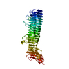
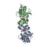
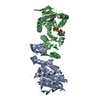
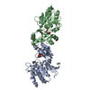
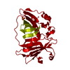
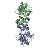
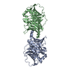
登録情報 データベース : PDB / ID : 3pb1タイトル Crystal Structure of a Michaelis Complex between Plasminogen Activator Inhibitor-1 and Urokinase-type Plasminogen Activator Plasminogen activator inhibitor 1 Plasminogen activator, urokinase キーワード / / / / / / / 機能・相同性 分子機能 ドメイン・相同性 構成要素
/ / / / / / / / / / / / / / / / / / / / / / / / / / / / / / / / / / / / / / / / / / / / / / / / / / / / / / / / / / / / / / / / / / / / / / / / / / / / / / / / / / / / / / / / / / / / / / / / / / / / / / / / / / / / / / / / / / / / / / / / / / / / / / / / / / / / / 生物種 Homo sapiens (ヒト)手法 / / / 解像度 : 2.3 Å データ登録者 Lin, Z. / Jiang, L. / Huang, M. / Structure 2 Function Project (S2F) ジャーナル : J.Biol.Chem. / 年 : 2011タイトル : Structural basis for recognition of urokinase-type plasminogen activator by plasminogen activator inhibitor-1.著者 : Lin, Z. / Jiang, L. / Yuan, C. / Jensen, J.K. / Zhang, X. / Luo, Z. / Furie, B.C. / Furie, B. / Andreasen, P.A. / Huang, M. 履歴 登録 2010年10月20日 登録サイト / 処理サイト 改定 1.0 2010年12月29日 Provider / タイプ 改定 1.1 2011年7月13日 Group 改定 1.2 2023年11月1日 Group Data collection / Database references ... Data collection / Database references / Derived calculations / Refinement description カテゴリ chem_comp_atom / chem_comp_bond ... chem_comp_atom / chem_comp_bond / database_2 / pdbx_initial_refinement_model / struct_ref_seq_dif / struct_site Item _database_2.pdbx_DOI / _database_2.pdbx_database_accession ... _database_2.pdbx_DOI / _database_2.pdbx_database_accession / _struct_ref_seq_dif.details / _struct_site.pdbx_auth_asym_id / _struct_site.pdbx_auth_comp_id / _struct_site.pdbx_auth_seq_id 改定 1.3 2024年11月6日 Group カテゴリ / pdbx_modification_feature
すべて表示 表示を減らす
 ムービー
ムービー コントローラー


 データを開く
 基本情報
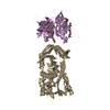
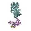
基本情報 要素
要素 キーワード
キーワード 機能・相同性情報
 Homo sapiens (ヒト)
 X線回折 /
  データ登録者
 引用
引用 構造の表示
構造の表示 ダウンロードとリンク
 その他のダウンロード


 PDBj
PDBj


 集合体
集合体

 Pichia pastoris (菌類)
 分子量: 96.063 Da / 分子数: 2 / 由来タイプ: 合成 / 式: SO4
 分子量: 18.015 Da / 分子数: 314 / 由来タイプ: 天然 / 式: H2O
 試料調製
試料調製 / ビームライン: BL17U / 波長: 1 Å
 解析
解析