 Movie
Movie Controller

[English] 日本語
 Yorodumi
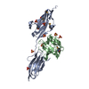
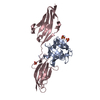
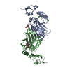
- PDB-3ojv: Crystal Structure of FGF1 complexed with the ectodomain of FGFR1c... -
+ Open data

- Basic information
| Entry | Database: PDB / ID: 3ojv | |||||||||
|---|---|---|---|---|---|---|---|---|---|---|
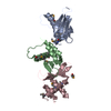
| Title | Crystal Structure of FGF1 complexed with the ectodomain of FGFR1c exhibiting an ordered ligand specificity-determining betaC'-betaE loop | |||||||||
 Components Components |
| |||||||||
 Keywords Keywords | Cytokine/Signaling Protein / beta trefoil motif / immunoglobulin-like domain / growth factor / growth factor receptor / extracellular / Cytokine-Signaling Protein complex | |||||||||
| Function / homology |  Function and homology information Function and homology informationSignaling by FGFR1 amplification mutants / negative regulation of fibroblast growth factor production / positive regulation of mitotic cell cycle DNA replication / regulation of extrinsic apoptotic signaling pathway in absence of ligand / diphosphate metabolic process / Signaling by plasma membrane FGFR1 fusions / FGFR1c and Klotho ligand binding and activation / regulation of lateral mesodermal cell fate specification / positive regulation of MAPKKK cascade by fibroblast growth factor receptor signaling pathway / mesonephric epithelium development ...Signaling by FGFR1 amplification mutants / negative regulation of fibroblast growth factor production / positive regulation of mitotic cell cycle DNA replication / regulation of extrinsic apoptotic signaling pathway in absence of ligand / diphosphate metabolic process / Signaling by plasma membrane FGFR1 fusions / FGFR1c and Klotho ligand binding and activation / regulation of lateral mesodermal cell fate specification / positive regulation of MAPKKK cascade by fibroblast growth factor receptor signaling pathway / mesonephric epithelium development / branch elongation involved in ureteric bud branching / vitamin D3 metabolic process / regulation of endothelial tube morphogenesis / regulation of phosphate transport / cementum mineralization / FGFR3b ligand binding and activation / regulation of endothelial cell chemotaxis to fibroblast growth factor / regulation of branching involved in salivary gland morphogenesis by mesenchymal-epithelial signaling / response to sodium phosphate / Epithelial-Mesenchymal Transition (EMT) during gastrulation / fibroblast growth factor receptor signaling pathway involved in orbitofrontal cortex development / ventricular zone neuroblast division / Signaling by activated point mutants of FGFR3 / FGFR3c ligand binding and activation / mesenchymal cell proliferation / Phospholipase C-mediated cascade; FGFR3 / positive regulation of phospholipase activity / receptor-receptor interaction / chordate embryonic development / positive regulation of parathyroid hormone secretion / fibroblast growth factor receptor binding / FGFR2b ligand binding and activation / auditory receptor cell development / paraxial mesoderm development / FGFR2c ligand binding and activation / Activated point mutants of FGFR2 / FGFR4 ligand binding and activation / Phospholipase C-mediated cascade; FGFR2 / regulation of postsynaptic density assembly / FGFR1b ligand binding and activation / Phospholipase C-mediated cascade; FGFR4 / Signaling by activated point mutants of FGFR1 / FGFR1c ligand binding and activation / organ induction / Downstream signaling of activated FGFR1 / Phospholipase C-mediated cascade: FGFR1 / fibroblast growth factor receptor activity / branching involved in salivary gland morphogenesis / lung-associated mesenchyme development / cell projection assembly / S100 protein binding / outer ear morphogenesis / activation of protein kinase B activity / embryonic limb morphogenesis / positive regulation of vascular endothelial cell proliferation / positive regulation of endothelial cell chemotaxis / ureteric bud development / positive regulation of mesenchymal cell proliferation / skeletal system morphogenesis / Signaling by FGFR2 IIIa TM / inner ear morphogenesis / PI-3K cascade:FGFR3 / middle ear morphogenesis / positive regulation of stem cell proliferation / Formation of paraxial mesoderm / PI-3K cascade:FGFR2 / PI-3K cascade:FGFR4 / positive regulation of sprouting angiogenesis / PI-3K cascade:FGFR1 / midbrain development / positive regulation of MAP kinase activity / phosphatidylinositol-mediated signaling / regulation of cell differentiation / positive regulation of cell division / positive regulation of intracellular signal transduction / fibroblast growth factor binding / fibroblast growth factor receptor signaling pathway / PI3K Cascade / epithelial to mesenchymal transition / positive regulation of blood vessel endothelial cell migration / chondrocyte differentiation / anatomical structure morphogenesis / cardiac muscle cell proliferation / positive regulation of cardiac muscle cell proliferation / SHC-mediated cascade:FGFR3 / SHC-mediated cascade:FGFR2 / SHC-mediated cascade:FGFR4 / calcium ion homeostasis / SHC-mediated cascade:FGFR1 / FRS-mediated FGFR3 signaling / FRS-mediated FGFR2 signaling / FRS-mediated FGFR4 signaling / peptidyl-tyrosine phosphorylation / FRS-mediated FGFR1 signaling / Signaling by FGFR3 in disease / cell maturation / Signaling by FGFR2 in disease / cellular response to fibroblast growth factor stimulus / positive regulation of neuron differentiation / Signaling by FGFR1 in disease Similarity search - Function | |||||||||
| Biological species |  Homo sapiens (human) Homo sapiens (human) | |||||||||
| Method |  X-RAY DIFFRACTION / SYNCHROTRON / MOLECULAR REPLACEMENT / Resolution: 2.6 Å X-RAY DIFFRACTION / SYNCHROTRON / MOLECULAR REPLACEMENT / Resolution: 2.6 Å | |||||||||
 Authors Authors | Beenken, A. / Mohammadi, M. | |||||||||
 Citation Citation | Journal: J.Biol.Chem. / Year: 2012 Title: Plasticity in Interactions of Fibroblast Growth Factor 1 (FGF1) N Terminus with FGF Receptors Underlies Promiscuity of FGF1. Authors: Beenken, A. / Eliseenkova, A.V. / Ibrahimi, O.A. / Olsen, S.K. / Mohammadi, M. | |||||||||
| History |
|
- Structure visualization
| Structure viewer | Molecule: MolmilJmol/JSmol |
|---|
- Downloads & links
-Download
| PDBx/mmCIF format | 3ojv.cif.gz | 149.2 KB | Display | PDBx/mmCIF format |
|---|---|---|---|---|
| PDB format | pdb3ojv.ent.gz | 115.1 KB | Display | PDB format |
| PDBx/mmJSON format | 3ojv.json.gz | Tree view | PDBx/mmJSON format | |
| Others |  Other downloads Other downloads |
-Validation report
| Arichive directory | https://data.pdbj.org/pub/pdb/validation_reports/oj/3ojvftp://data.pdbj.org/pub/pdb/validation_reports/oj/3ojv | HTTPS FTP |
|---|
-Related structure data
| Related structure data |  3oj2C  3ojmC 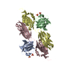 1evtS S: Starting model for refinement C: citing same article ( |
|---|---|
| Similar structure data |


-Links
 PDBj
PDBj


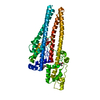
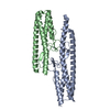
- Assembly
| Deposited unit | 
| ||||||||
|---|---|---|---|---|---|---|---|---|---|
| 1 | 
| ||||||||
| 2 | 
| ||||||||
| Unit cell |
|
-Components
| #1: Protein | Mass: 15420.397 Da / Num. of mol.: 2 Source method: isolated from a genetically manipulated source Source: (gene. exp.) Homo sapiens (human) / Gene: FGF1, FGFA / Production host:  #2: Protein | Mass: 25375.973 Da / Num. of mol.: 2 / Fragment: FGFR1c / Mutation: N185Q Source method: isolated from a genetically manipulated source Source: (gene. exp.) Homo sapiens (human) / Gene: FGFBR, FGFR1, FLG, FLT2 / Plasmid: pET28a / Production host: References: UniProt: P11362, receptor protein-tyrosine kinase #3: Polysaccharide | 4-deoxy-2-O-sulfo-alpha-L-threo-hex-4-enopyranuronic acid-(1-4)-2-deoxy-6-O-sulfo-2-(sulfoamino)- ...4-deoxy-2-O-sulfo-alpha-L-threo-hex-4-enopyranuronic acid-(1-4)-2-deoxy-6-O-sulfo-2-(sulfoamino)-alpha-D-glucopyranose-(1-4)-2-O-sulfo-alpha-L-idopyranuronic acid-(1-4)-2-deoxy-6-O-sulfo-2-(sulfoamino)-alpha-D-glucopyranose-(1-4)-2-O-sulfo-alpha-L-idopyranuronic acid-(1-4)-2-deoxy-6-O-sulfo-2-(sulfoamino)-alpha-D-glucopyranose | Source method: isolated from a genetically manipulated source #4: Water | ChemComp-HOH / |  Mass: 18.015 Da / Num. of mol.: 32 / Source method: isolated from a natural source / Formula: H2O Mass: 18.015 Da / Num. of mol.: 32 / Source method: isolated from a natural source / Formula: H2OHas protein modification | Y | |
|---|
-Experimental details
-Experiment
| Experiment | Method: X-RAY DIFFRACTION / Number of used crystals: 1 |
|---|
- Sample preparation
| Crystal | Density Matthews: 2.55 Å3/Da / Density % sol: 51.7 % |
|---|---|
| Crystal grow | Temperature: 293 K / Method: vapor diffusion, hanging drop / pH: 8.5 Details: 0.1M Tris, 15% PEG4000, 0.1M ammonium sulfate, pH 8.5, VAPOR DIFFUSION, HANGING DROP, temperature 293K |
-Data collection
| Diffraction | Mean temperature: 173 K | ||||||||||||||||||||||||||||||||||||||||||||||||||||||||||||||||||
|---|---|---|---|---|---|---|---|---|---|---|---|---|---|---|---|---|---|---|---|---|---|---|---|---|---|---|---|---|---|---|---|---|---|---|---|---|---|---|---|---|---|---|---|---|---|---|---|---|---|---|---|---|---|---|---|---|---|---|---|---|---|---|---|---|---|---|---|
| Diffraction source | Source: SYNCHROTRON / Site: NSLS  / Beamline: X4A / Wavelength: 0.97912 Å / Beamline: X4A / Wavelength: 0.97912 Å | ||||||||||||||||||||||||||||||||||||||||||||||||||||||||||||||||||
| Detector | Type: ADSC QUANTUM 4 / Detector: CCD / Date: Feb 26, 2005 | ||||||||||||||||||||||||||||||||||||||||||||||||||||||||||||||||||
| Radiation | Monochromator: KOHZU DOUBLE CRYSTAL / Protocol: SINGLE WAVELENGTH / Monochromatic (M) / Laue (L): M / Scattering type: x-ray | ||||||||||||||||||||||||||||||||||||||||||||||||||||||||||||||||||
| Radiation wavelength | Wavelength: 0.97912 Å / Relative weight: 1 | ||||||||||||||||||||||||||||||||||||||||||||||||||||||||||||||||||
| Reflection | Resolution: 2.6→50 Å / Num. all: 24626 / Num. obs: 24626 / % possible obs: 97.8 % / Observed criterion σ(F): 0 / Observed criterion σ(I): 0 / Redundancy: 3.7 % / Rsym value: 0.064 / Net I/σ(I): 16.6 | ||||||||||||||||||||||||||||||||||||||||||||||||||||||||||||||||||
| Reflection shell |
|
- Processing
| Software |
| ||||||||||||||||||||||||||||||||||||||||||||||||||||||||||||
|---|---|---|---|---|---|---|---|---|---|---|---|---|---|---|---|---|---|---|---|---|---|---|---|---|---|---|---|---|---|---|---|---|---|---|---|---|---|---|---|---|---|---|---|---|---|---|---|---|---|---|---|---|---|---|---|---|---|---|---|---|---|
| Refinement | Method to determine structure: MOLECULAR REPLACEMENT Starting model: PDB entry 1EVT Resolution: 2.6→25 Å / Cross valid method: THROUGHOUT / σ(F): 0 / σ(I): 0 / Stereochemistry target values: Engh & Huber Details: There is substantial electron density in the difference Fourier synthesis (Fo - Fc) map contoured at 1 sigma level between the heparin binding sites of the two FGFs. The shape of the ...Details: There is substantial electron density in the difference Fourier synthesis (Fo - Fc) map contoured at 1 sigma level between the heparin binding sites of the two FGFs. The shape of the electron density and its location (sandwiched between heparin binding sites of the two FGFs) clearly indicate that it belongs to the cocrystallized heparin octasaccharide. However, only six monosaccharides of the octasacchride could be confidently built into this density. Importantly, there are densities of the size of a disaccharide unit at either end of this modeled hexasaccharide suggesting that the hexasaccharide could be translated by two sugar units along its helical axis. This observation suggests that the octasaccharide does not bind in a fixed fashion to the FGFs and can glide by two sugar units along its axis. As a result, the temperature factor and the real space R factor for the octasaccharide are abnormally high.
| ||||||||||||||||||||||||||||||||||||||||||||||||||||||||||||
| Displacement parameters |
| ||||||||||||||||||||||||||||||||||||||||||||||||||||||||||||
| Refinement step | Cycle: LAST / Resolution: 2.6→25 Å
| ||||||||||||||||||||||||||||||||||||||||||||||||||||||||||||
| Refine LS restraints |
| ||||||||||||||||||||||||||||||||||||||||||||||||||||||||||||
| LS refinement shell |
|
