 Movie
Movie Controller

[English] 日本語
 Yorodumi
- PDB-1evt: CRYSTAL STRUCTURE OF FGF1 IN COMPLEX WITH THE EXTRACELLULAR LIGAN... -
+ Open data

- Basic information
| Entry | Database: PDB / ID: 1evt | ||||||
|---|---|---|---|---|---|---|---|
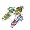
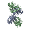
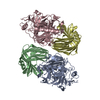
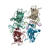
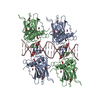
| Title | CRYSTAL STRUCTURE OF FGF1 IN COMPLEX WITH THE EXTRACELLULAR LIGAND BINDING DOMAIN OF FGF RECEPTOR 1 (FGFR1) | ||||||
 Components Components |
| ||||||
 Keywords Keywords | GROWTH FACTOR/GROWTH FACTOR RECEPTOR / IMMUNOGLOBULIN (IG) LIKE DOMAINS BELONGING TO THE I-SET SUBGROUP WITHIN IG-LIKE DOMAINS / B-TREFOIL FOLD / GROWTH FACTOR-GROWTH FACTOR RECEPTOR COMPLEX | ||||||
| Function / homology |  Function and homology information Function and homology informationSignaling by FGFR1 amplification mutants / negative regulation of fibroblast growth factor production / positive regulation of mitotic cell cycle DNA replication / regulation of extrinsic apoptotic signaling pathway in absence of ligand / diphosphate metabolic process / Signaling by plasma membrane FGFR1 fusions / FGFR1c and Klotho ligand binding and activation / regulation of lateral mesodermal cell fate specification / positive regulation of MAPKKK cascade by fibroblast growth factor receptor signaling pathway / regulation of phosphate transport ...Signaling by FGFR1 amplification mutants / negative regulation of fibroblast growth factor production / positive regulation of mitotic cell cycle DNA replication / regulation of extrinsic apoptotic signaling pathway in absence of ligand / diphosphate metabolic process / Signaling by plasma membrane FGFR1 fusions / FGFR1c and Klotho ligand binding and activation / regulation of lateral mesodermal cell fate specification / positive regulation of MAPKKK cascade by fibroblast growth factor receptor signaling pathway / regulation of phosphate transport / mesonephric epithelium development / branch elongation involved in ureteric bud branching / vitamin D3 metabolic process / regulation of endothelial tube morphogenesis / cementum mineralization / FGFR3b ligand binding and activation / regulation of branching involved in salivary gland morphogenesis by mesenchymal-epithelial signaling / regulation of endothelial cell chemotaxis to fibroblast growth factor / response to sodium phosphate / Epithelial-Mesenchymal Transition (EMT) during gastrulation / fibroblast growth factor receptor signaling pathway involved in orbitofrontal cortex development / ventricular zone neuroblast division / Signaling by activated point mutants of FGFR3 / FGFR3c ligand binding and activation / mesenchymal cell proliferation / Phospholipase C-mediated cascade; FGFR3 / positive regulation of phospholipase activity / receptor-receptor interaction / chordate embryonic development / positive regulation of parathyroid hormone secretion / fibroblast growth factor receptor binding / FGFR2b ligand binding and activation / auditory receptor cell development / paraxial mesoderm development / FGFR2c ligand binding and activation / Activated point mutants of FGFR2 / FGFR4 ligand binding and activation / Phospholipase C-mediated cascade; FGFR2 / regulation of postsynaptic density assembly / FGFR1b ligand binding and activation / Phospholipase C-mediated cascade; FGFR4 / organ induction / Signaling by activated point mutants of FGFR1 / FGFR1c ligand binding and activation / Downstream signaling of activated FGFR1 / fibroblast growth factor receptor activity / Phospholipase C-mediated cascade: FGFR1 / branching involved in salivary gland morphogenesis / lung-associated mesenchyme development / cell projection assembly / S100 protein binding / outer ear morphogenesis / activation of protein kinase B activity / embryonic limb morphogenesis / positive regulation of vascular endothelial cell proliferation / positive regulation of endothelial cell chemotaxis / ureteric bud development / positive regulation of mesenchymal cell proliferation / skeletal system morphogenesis / Signaling by FGFR2 IIIa TM / inner ear morphogenesis / PI-3K cascade:FGFR3 / middle ear morphogenesis / Formation of paraxial mesoderm / PI-3K cascade:FGFR2 / positive regulation of stem cell proliferation / PI-3K cascade:FGFR4 / positive regulation of sprouting angiogenesis / PI-3K cascade:FGFR1 / midbrain development / positive regulation of MAP kinase activity / phosphatidylinositol-mediated signaling / regulation of cell differentiation / positive regulation of cell division / positive regulation of intracellular signal transduction / fibroblast growth factor binding / fibroblast growth factor receptor signaling pathway / PI3K Cascade / epithelial to mesenchymal transition / positive regulation of blood vessel endothelial cell migration / chondrocyte differentiation / anatomical structure morphogenesis / cardiac muscle cell proliferation / positive regulation of cardiac muscle cell proliferation / SHC-mediated cascade:FGFR3 / SHC-mediated cascade:FGFR2 / calcium ion homeostasis / SHC-mediated cascade:FGFR4 / SHC-mediated cascade:FGFR1 / FRS-mediated FGFR3 signaling / FRS-mediated FGFR2 signaling / FRS-mediated FGFR4 signaling / peptidyl-tyrosine phosphorylation / FRS-mediated FGFR1 signaling / Signaling by FGFR3 in disease / cell maturation / cellular response to fibroblast growth factor stimulus / Signaling by FGFR2 in disease / positive regulation of neuron differentiation / Signaling by FGFR1 in disease Similarity search - Function | ||||||
| Biological species |  Homo sapiens (human) Homo sapiens (human) | ||||||
| Method |  X-RAY DIFFRACTION / SYNCHROTRON / Resolution: 2.8 Å X-RAY DIFFRACTION / SYNCHROTRON / Resolution: 2.8 Å | ||||||
 Authors Authors | Plotnikov, A.N. / Hubbard, S.R. / Schlessinger, J. / Mohammadi, M. | ||||||
 Citation Citation | Journal: Cell(Cambridge,Mass.) / Year: 2000 Title: Crystal structures of two FGF-FGFR complexes reveal the determinants of ligand-receptor specificity. Authors: Plotnikov, A.N. / Hubbard, S.R. / Schlessinger, J. / Mohammadi, M. | ||||||
| History |
|
- Structure visualization
| Structure viewer | Molecule: MolmilJmol/JSmol |
|---|
- Downloads & links
-Download
| PDBx/mmCIF format | 1evt.cif.gz | 135 KB | Display | PDBx/mmCIF format |
|---|---|---|---|---|
| PDB format | pdb1evt.ent.gz | 104.8 KB | Display | PDB format |
| PDBx/mmJSON format | 1evt.json.gz | Tree view | PDBx/mmJSON format | |
| Others |  Other downloads Other downloads |
-Validation report
| Arichive directory | https://data.pdbj.org/pub/pdb/validation_reports/ev/1evtftp://data.pdbj.org/pub/pdb/validation_reports/ev/1evt | HTTPS FTP |
|---|
-Related structure data


-Links
 PDBj
PDBj


- Assembly
| Deposited unit | 
| ||||||||
|---|---|---|---|---|---|---|---|---|---|
| 1 |
| ||||||||
| Unit cell |
|
-Components
| #1: Protein | Mass: 15232.148 Da / Num. of mol.: 2 Fragment: THE B-TREFOIL CORE OF FIBROBLAST GROWTH FACTOR 1 (FGF1) Source method: isolated from a genetically manipulated source Source: (gene. exp.) Homo sapiens (human) / Production host: Bacteria (eubacteria) / References: UniProt: P05230#2: Protein | Mass: 25260.777 Da / Num. of mol.: 2 Fragment: EXTRACELLULAR LIGAND BINDING DOMAIN OF FGF RECEPTOR 1 (FGFR1) CONSISTING OF IMMUNOGLOBULIN LIKE DOMAINS II (D2) AND III (D3) Source method: isolated from a genetically manipulated source Source: (gene. exp.) Homo sapiens (human) / Production host: Bacteria (eubacteria) / References: UniProt: P11362#3: Chemical | ChemComp-SO4 /   Mass: 96.063 Da / Num. of mol.: 4 / Source method: obtained synthetically / Formula: SO4 Mass: 96.063 Da / Num. of mol.: 4 / Source method: obtained synthetically / Formula: SO4Has protein modification | Y | |
|---|
-Experimental details
-Experiment
| Experiment | Method: X-RAY DIFFRACTION / Number of used crystals: 1 |
|---|
- Sample preparation
| Crystal | Density Matthews: 2.92 Å3/Da / Density % sol: 57.82 % | ||||||||||||||||||||||||||||||||||||
|---|---|---|---|---|---|---|---|---|---|---|---|---|---|---|---|---|---|---|---|---|---|---|---|---|---|---|---|---|---|---|---|---|---|---|---|---|---|
| Crystal grow | Temperature: 298 K / Method: vapor diffusion / pH: 7.5 Details: PEG 4000, Isopropanol, HEPES-NaOH, pH 7.5, VAPOR DIFFUSION, temperature 298.0K | ||||||||||||||||||||||||||||||||||||
| Crystal grow | *PLUS Temperature: 20 ℃ / Method: vapor diffusion, hanging drop | ||||||||||||||||||||||||||||||||||||
| Components of the solutions | *PLUS
|
-Data collection
| Diffraction | Mean temperature: 110 K |
|---|---|
| Diffraction source | Source: SYNCHROTRON / Site: NSLS  / Beamline: X4A / Wavelength: 0.9794 / Beamline: X4A / Wavelength: 0.9794 |
| Detector | Type: RIGAKU RAXIS IV / Detector: IMAGE PLATE / Date: Mar 25, 1999 |
| Radiation | Protocol: SINGLE WAVELENGTH / Monochromatic (M) / Laue (L): M / Scattering type: x-ray |
| Radiation wavelength | Wavelength: 0.9794 Å / Relative weight: 1 |
| Reflection | Resolution: 2.8→25 Å / Num. obs: 49288 / % possible obs: 97.9 % / Observed criterion σ(I): 0 / Redundancy: 2.2 % / Rmerge(I) obs: 0.083 / Net I/σ(I): 8.6 |
| Reflection shell | Resolution: 2.8→2.9 Å / Rmerge(I) obs: 0.226 / % possible all: 90.5 |
| Reflection | *PLUS Num. obs: 22330 / Num. measured all: 49288 |
| Reflection shell | *PLUS % possible obs: 90.5 % |
- Processing
| Software |
| ||||||||||||||||||||||||||||||||||||||||||||||||||||||||||||||||||||||||||||||||
|---|---|---|---|---|---|---|---|---|---|---|---|---|---|---|---|---|---|---|---|---|---|---|---|---|---|---|---|---|---|---|---|---|---|---|---|---|---|---|---|---|---|---|---|---|---|---|---|---|---|---|---|---|---|---|---|---|---|---|---|---|---|---|---|---|---|---|---|---|---|---|---|---|---|---|---|---|---|---|---|---|---|
| Refinement | Resolution: 2.8→25 Å / σ(F): 0 / Stereochemistry target values: ENGH & HUBER
| ||||||||||||||||||||||||||||||||||||||||||||||||||||||||||||||||||||||||||||||||
| Solvent computation | Solvent model: CNS / Bsol: 27.76 Å2 / ksol: 0.342 e/Å3 | ||||||||||||||||||||||||||||||||||||||||||||||||||||||||||||||||||||||||||||||||
| Displacement parameters |
| ||||||||||||||||||||||||||||||||||||||||||||||||||||||||||||||||||||||||||||||||
| Refinement step | Cycle: LAST / Resolution: 2.8→25 Å
| ||||||||||||||||||||||||||||||||||||||||||||||||||||||||||||||||||||||||||||||||
| Refine LS restraints |
| ||||||||||||||||||||||||||||||||||||||||||||||||||||||||||||||||||||||||||||||||
| Software | *PLUS Name: 'CNS' / Classification: refinement | ||||||||||||||||||||||||||||||||||||||||||||||||||||||||||||||||||||||||||||||||
| Refine LS restraints | *PLUS
|
