 Movie
Movie Controller

[English] 日本語
 Yorodumi
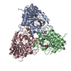
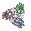
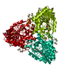
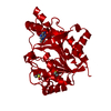
- PDB-3odg: crystal structure of xanthosine phosphorylase bound with xanthine... -
+ Open data

- Basic information
| Entry | Database: PDB / ID: 3odg | ||||||
|---|---|---|---|---|---|---|---|
| Title | crystal structure of xanthosine phosphorylase bound with xanthine from Yersinia pseudotuberculosis | ||||||
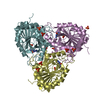
 Components Components | Xanthosine phosphorylase | ||||||
 Keywords Keywords | TRANSFERASE / Structural Genomics / PSI-2 / Protein Structure Initiative / New York SGX Research Center for Structural Genomics / NYSGXRC / Purine Nucleoside Phosphorylase / purine nucleoside binding | ||||||
| Function / homology |  Function and homology information Function and homology informationnucleoside metabolic process / purine-nucleoside phosphorylase activity / purine-nucleoside phosphorylase / cytoplasm Similarity search - Function | ||||||
| Biological species |  Yersinia pseudotuberculosis (bacteria) Yersinia pseudotuberculosis (bacteria) | ||||||
| Method |  X-RAY DIFFRACTION / SYNCHROTRON / MOLECULAR REPLACEMENT / Resolution: 1.64 Å X-RAY DIFFRACTION / SYNCHROTRON / MOLECULAR REPLACEMENT / Resolution: 1.64 Å | ||||||
 Authors Authors | Kim, J. / Ramagopal, U.A. / Burley, S.K. / Almo, S.C. / New York SGX Research Center for Structural Genomics (NYSGXRC) | ||||||
 Citation Citation | Journal: To be Published Title: crystal structure of xanthosine phosphorylase bound with xanthine from Yersinia pseudotuberculosis Authors: Kim, J. / Ramagopal, U.A. / Almo, S.C. | ||||||
| History |
|
- Structure visualization
| Structure viewer | Molecule: MolmilJmol/JSmol |
|---|
- Downloads & links
-Download
| PDBx/mmCIF format | 3odg.cif.gz | 125.1 KB | Display | PDBx/mmCIF format |
|---|---|---|---|---|
| PDB format | pdb3odg.ent.gz | 97.4 KB | Display | PDB format |
| PDBx/mmJSON format | 3odg.json.gz | Tree view | PDBx/mmJSON format | |
| Others |  Other downloads Other downloads |
-Validation report
| Arichive directory | https://data.pdbj.org/pub/pdb/validation_reports/od/3odgftp://data.pdbj.org/pub/pdb/validation_reports/od/3odg | HTTPS FTP |
|---|
-Related structure data
| Related structure data |  1yqqS S: Starting model for refinement |
|---|---|
| Similar structure data |


-Links
 PDBj
PDBj
- Assembly
| Deposited unit | 
| ||||||||
|---|---|---|---|---|---|---|---|---|---|
| 1 | 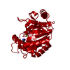
| ||||||||
| Unit cell |
|
-Components
| #1: Protein | Mass: 30942.965 Da / Num. of mol.: 1 Source method: isolated from a genetically manipulated source Source: (gene. exp.) Yersinia pseudotuberculosis (bacteria) / Gene: pndA, xapA, YPTB1201 / Plasmid: LIC-PET30a / Production host: References: UniProt: Q66D48, Transferases; Glycosyltransferases; Pentosyltransferases |
|---|---|
| #2: Chemical | ChemComp-XAN / 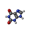  Mass: 152.111 Da / Num. of mol.: 1 / Source method: obtained synthetically / Formula: C5H4N4O2 Mass: 152.111 Da / Num. of mol.: 1 / Source method: obtained synthetically / Formula: C5H4N4O2 |
| #3: Chemical | ChemComp-CL /   Mass: 35.453 Da / Num. of mol.: 1 / Source method: obtained synthetically / Formula: Cl Mass: 35.453 Da / Num. of mol.: 1 / Source method: obtained synthetically / Formula: Cl |
| #4: Water | ChemComp-HOH /  Mass: 18.015 Da / Num. of mol.: 252 / Source method: isolated from a natural source / Formula: H2O Mass: 18.015 Da / Num. of mol.: 252 / Source method: isolated from a natural source / Formula: H2O |
-Experimental details
-Experiment
| Experiment | Method: X-RAY DIFFRACTION / Number of used crystals: 1 |
|---|
- Sample preparation
| Crystal | Density Matthews: 2.12 Å3/Da / Density % sol: 42.06 % |
|---|---|
| Crystal grow | Temperature: 293 K / Method: vapor diffusion, sitting drop / pH: 8.5 Details: 0.1M Tris-HCl PH 8.5, 0.2M Sodium Acetate, 30% PEG4000, VAPOR DIFFUSION, SITTING DROP, temperature 293K |
-Data collection
| Diffraction | Mean temperature: 100 K |
|---|---|
| Diffraction source | Source: SYNCHROTRON / Site: NSLS  / Beamline: X29A / Wavelength: 1.075 Å / Beamline: X29A / Wavelength: 1.075 Å |
| Detector | Type: ADSC QUANTUM 315 / Detector: CCD / Date: Oct 9, 2009 |
| Radiation | Monochromator: Double silicon(111) crystal / Protocol: SINGLE WAVELENGTH / Monochromatic (M) / Laue (L): M / Scattering type: x-ray |
| Radiation wavelength | Wavelength: 1.075 Å / Relative weight: 1 |
| Reflection | Resolution: 1.64→48.24 Å / Num. all: 31965 / Num. obs: 31056 / % possible obs: 97.2 % / Redundancy: 11.7 % / Biso Wilson estimate: 26.5 Å2 / Rmerge(I) obs: 0.096 / Rsym value: 0.076 / Net I/σ(I): 24.7 |
| Reflection shell | Resolution: 1.64→1.7 Å / Redundancy: 10.2 % / Rmerge(I) obs: 0.562 / Mean I/σ(I) obs: 3.1 / Num. unique all: 2550 / Rsym value: 0.483 / % possible all: 81.1 |
- Processing
| Software |
| |||||||||||||||||||||||||||||||||||||||||||||||||||||||||||||||||
|---|---|---|---|---|---|---|---|---|---|---|---|---|---|---|---|---|---|---|---|---|---|---|---|---|---|---|---|---|---|---|---|---|---|---|---|---|---|---|---|---|---|---|---|---|---|---|---|---|---|---|---|---|---|---|---|---|---|---|---|---|---|---|---|---|---|---|
| Refinement | Method to determine structure: MOLECULAR REPLACEMENT Starting model: 1YQQ Resolution: 1.64→48.24 Å / Cor.coef. Fo:Fc: 0.973 / Cor.coef. Fo:Fc free: 0.964 / SU B: 4.259 / SU ML: 0.065 / Cross valid method: THROUGHOUT / σ(F): 1 / ESU R Free: 0.089 / Stereochemistry target values: MAXIMUM LIKELIHOOD / Details: HYDROGENS HAVE BEEN ADDED IN THE RIDING POSITIONS
| |||||||||||||||||||||||||||||||||||||||||||||||||||||||||||||||||
| Solvent computation | Ion probe radii: 0.8 Å / Shrinkage radii: 0.8 Å / VDW probe radii: 1.4 Å / Solvent model: MASK | |||||||||||||||||||||||||||||||||||||||||||||||||||||||||||||||||
| Displacement parameters | Biso mean: 24.427 Å2
| |||||||||||||||||||||||||||||||||||||||||||||||||||||||||||||||||
| Refinement step | Cycle: LAST / Resolution: 1.64→48.24 Å
| |||||||||||||||||||||||||||||||||||||||||||||||||||||||||||||||||
| Refine LS restraints |
| |||||||||||||||||||||||||||||||||||||||||||||||||||||||||||||||||
| LS refinement shell | Resolution: 1.639→1.682 Å / Total num. of bins used: 20
| |||||||||||||||||||||||||||||||||||||||||||||||||||||||||||||||||
| Refinement TLS params. | Method: refined / Origin x: -28.102 Å / Origin y: -19.958 Å / Origin z: -0.107 Å
|
