 Movie
Movie Controller

[English] 日本語
 Yorodumi
- PDB-3ml0: Thermostable Penicillin G acylase from Alcaligenes faecalis in te... -
+ Open data

- Basic information
| Entry | Database: PDB / ID: 3ml0 | ||||||
|---|---|---|---|---|---|---|---|
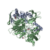
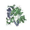
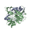
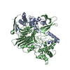
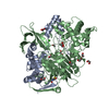
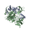
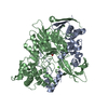
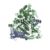
| Title | Thermostable Penicillin G acylase from Alcaligenes faecalis in tetragonal form | ||||||
 Components Components |
| ||||||
 Keywords Keywords | HYDROLASE / penicillin G acylase | ||||||
| Function / homology |  Function and homology information Function and homology informationpenicillin amidase activity / penicillin amidase / antibiotic biosynthetic process / metal ion binding Similarity search - Function | ||||||
| Biological species |  Alcaligenes faecalis (bacteria) Alcaligenes faecalis (bacteria) | ||||||
| Method |  X-RAY DIFFRACTION / MOLECULAR REPLACEMENT / molecular replacement / Resolution: 3.5 Å X-RAY DIFFRACTION / MOLECULAR REPLACEMENT / molecular replacement / Resolution: 3.5 Å | ||||||
 Authors Authors | Varshney, N.K. / Kumar, R.S. / Ignatova, Z. / Dodson, E. / Suresh, C.G. | ||||||
 Citation Citation | Journal: Acta Crystallogr.,Sect.F / Year: 2012 Title: Crystallization and X-ray structure analysis of a thermostable penicillin G acylase from Alcaligenes faecalis. Authors: Varshney, N.K. / Kumar, R.S. / Ignatova, Z. / Prabhune, A. / Pundle, A. / Dodson, E. / Suresh, C.G. | ||||||
| History |
|
- Structure visualization
| Structure viewer | Molecule: MolmilJmol/JSmol |
|---|
- Downloads & links
-Download
| PDBx/mmCIF format | 3ml0.cif.gz | 152.7 KB | Display | PDBx/mmCIF format |
|---|---|---|---|---|
| PDB format | pdb3ml0.ent.gz | 119.9 KB | Display | PDB format |
| PDBx/mmJSON format | 3ml0.json.gz | Tree view | PDBx/mmJSON format | |
| Others |  Other downloads Other downloads |
-Validation report
| Arichive directory | https://data.pdbj.org/pub/pdb/validation_reports/ml/3ml0ftp://data.pdbj.org/pub/pdb/validation_reports/ml/3ml0 | HTTPS FTP |
|---|
-Related structure data
| Related structure data |  3k3wC 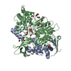 1gk9S C: citing same article ( S: Starting model for refinement |
|---|---|
| Similar structure data |


-Links
 PDBj
PDBj


- Assembly
| Deposited unit | 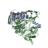
| ||||||||
|---|---|---|---|---|---|---|---|---|---|
| 1 |
| ||||||||
| Unit cell |
|
-Components
| #1: Protein | Mass: 22258.885 Da / Num. of mol.: 1 Source method: isolated from a genetically manipulated source Source: (gene. exp.) Alcaligenes faecalis (bacteria) / Gene: pac / Plasmid: pPAAF / Production host: |
|---|---|
| #2: Protein | Mass: 62791.953 Da / Num. of mol.: 1 Source method: isolated from a genetically manipulated source Source: (gene. exp.) Alcaligenes faecalis (bacteria) / Gene: pac / Plasmid: pPAAF / Production host: |
| #3: Chemical | ChemComp-CA /   Mass: 40.078 Da / Num. of mol.: 1 / Source method: obtained synthetically / Formula: Ca Mass: 40.078 Da / Num. of mol.: 1 / Source method: obtained synthetically / Formula: Ca |
| Has protein modification | Y |
-Experimental details
-Experiment
| Experiment | Method: X-RAY DIFFRACTION / Number of used crystals: 1 |
|---|
- Sample preparation
| Crystal | Density Matthews: 3.22 Å3/Da / Density % sol: 61.74 % |
|---|---|
| Crystal grow | Temperature: 303 K / Method: vapor diffusion, hanging drop / pH: 7.5 Details: 15% PEG 8000, 0.1M Tris-Hcl pH7.5, 100uL b-octyl-glucopyranoside (0.50% w/v), vapor diffusion, hanging drop, temperature 303K |
-Data collection
| Diffraction | Mean temperature: 295 K |
|---|---|
| Diffraction source | Source: ROTATING ANODE / Type: RIGAKU / Wavelength: 1.514 Å |
| Detector | Type: RIGAKU RAXIS IV++ / Detector: IMAGE PLATE / Date: Apr 7, 2006 / Details: Mirrors |
| Radiation | Monochromator: Osmic mirror / Protocol: SINGLE WAVELENGTH / Monochromatic (M) / Laue (L): M / Scattering type: x-ray |
| Radiation wavelength | Wavelength: 1.514 Å / Relative weight: 1 |
| Reflection | Resolution: 3.5→82.2 Å / Num. obs: 14860 / % possible obs: 90.8 % / Observed criterion σ(F): 1 / Redundancy: 2.25 % / Biso Wilson estimate: 29.99 Å2 / Rmerge(I) obs: 0.099 / Net I/σ(I): 7.42 |
| Reflection shell | Resolution: 3.499→3.59 Å / Redundancy: 2.01 % / Rmerge(I) obs: 0.239 / Mean I/σ(I) obs: 3.08 / Num. unique all: 13376 / % possible all: 89.67 |
-Phasing
| Phasing | Method: molecular replacement |
|---|
- Processing
| Software |
| |||||||||||||||||||||||||||||||||||||||||||||||||||||||||||||||||
|---|---|---|---|---|---|---|---|---|---|---|---|---|---|---|---|---|---|---|---|---|---|---|---|---|---|---|---|---|---|---|---|---|---|---|---|---|---|---|---|---|---|---|---|---|---|---|---|---|---|---|---|---|---|---|---|---|---|---|---|---|---|---|---|---|---|---|
| Refinement | Method to determine structure: MOLECULAR REPLACEMENT Starting model: 1GK9 Resolution: 3.5→20 Å / Cor.coef. Fo:Fc: 0.849 / Cor.coef. Fo:Fc free: 0.823 / Occupancy max: 1 / Occupancy min: 0 / SU B: 35.918 / SU ML: 0.547 / Cross valid method: THROUGHOUT / σ(F): 1 / ESU R Free: 0.716 / Stereochemistry target values: MAXIMUM LIKELIHOOD Details: HYDROGENS HAVE BEEN ADDED IN THE RIDING POSITIONS U VALUES: REFINED INDIVIDUALLY
| |||||||||||||||||||||||||||||||||||||||||||||||||||||||||||||||||
| Solvent computation | Ion probe radii: 0.8 Å / Shrinkage radii: 0.8 Å / VDW probe radii: 1.4 Å / Solvent model: MASK | |||||||||||||||||||||||||||||||||||||||||||||||||||||||||||||||||
| Displacement parameters | Biso max: 65.2 Å2 / Biso mean: 47.781 Å2 / Biso min: 20 Å2
| |||||||||||||||||||||||||||||||||||||||||||||||||||||||||||||||||
| Refinement step | Cycle: LAST / Resolution: 3.5→20 Å
| |||||||||||||||||||||||||||||||||||||||||||||||||||||||||||||||||
| Refine LS restraints |
| |||||||||||||||||||||||||||||||||||||||||||||||||||||||||||||||||
| LS refinement shell | Resolution: 3.499→3.59 Å / Total num. of bins used: 20
|
