 Movie
Movie Controller


+ Open data

- Basic information
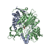
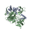
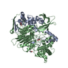
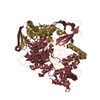
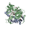
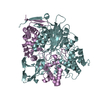
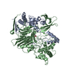
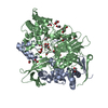
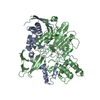
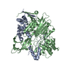
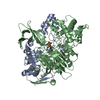
| Entry | Database: PDB / ID: 1k5s | ||||||
|---|---|---|---|---|---|---|---|
| Title | PENICILLIN ACYLASE, MUTANT COMPLEXED WITH PPA | ||||||
 Components Components |
| ||||||
 Keywords Keywords | HYDROLASE / NTN-HYDROLASE FOLD / HELICES / BETA-STRANDS | ||||||
| Function / homology |  Function and homology information Function and homology informationpenicillin amidase activity / penicillin amidase / antibiotic biosynthetic process / periplasmic space / response to antibiotic / metal ion binding Similarity search - Function | ||||||
| Biological species |  | ||||||
| Method |  X-RAY DIFFRACTION / MOLECULAR REPLACEMENT / Resolution: 2.43 Å X-RAY DIFFRACTION / MOLECULAR REPLACEMENT / Resolution: 2.43 Å | ||||||
 Authors Authors | Hensgens, C.M.H. / Keizer, E. / Snijder, H.J. / Dijkstra, B.W. | ||||||
 Citation Citation | Journal: Protein Eng.Des.Sel. / Year: 2004 Title: Structural and kinetic studies on ligand binding in wild-type and active-site mutants of penicillin acylase. Authors: Alkema, W.B.L. / Hensgens, C.M.H. / Snijder, H.J. / Keizer, E. / Dijkstra, B.W. / Janssen, D.B. | ||||||
| History |
|
- Structure visualization
| Structure viewer | Molecule: MolmilJmol/JSmol |
|---|
- Downloads & links
-Download
| PDBx/mmCIF format | 1k5s.cif.gz | 170.6 KB | Display | PDBx/mmCIF format |
|---|---|---|---|---|
| PDB format | pdb1k5s.ent.gz | 130.3 KB | Display | PDB format |
| PDBx/mmJSON format | 1k5s.json.gz | Tree view | PDBx/mmJSON format | |
| Others |  Other downloads Other downloads |
-Validation report
| Arichive directory | https://data.pdbj.org/pub/pdb/validation_reports/k5/1k5sftp://data.pdbj.org/pub/pdb/validation_reports/k5/1k5s | HTTPS FTP |
|---|
-Related structure data
| Related structure data |  1jx9C  1k5qC  1k7dC  1kecC 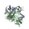 1pnkS C: citing same article ( S: Starting model for refinement |
|---|---|
| Similar structure data |


-Links
 PDBj
PDBj


- Assembly
| Deposited unit | 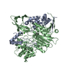
| ||||||||
|---|---|---|---|---|---|---|---|---|---|
| 1 |
| ||||||||
| Unit cell |
| ||||||||
| Details | The alpha and beta unit together form the biological unit / the alpha and beta unit together form the biological relevant unit |
-Components
| #1: Protein | Mass: 23838.822 Da / Num. of mol.: 1 Source method: isolated from a genetically manipulated source Source: (gene. exp.) |
|---|---|
| #2: Protein | Mass: 62367.426 Da / Num. of mol.: 1 / Mutation: F24A, V148L Source method: isolated from a genetically manipulated source Source: (gene. exp.) |
| #3: Chemical | ChemComp-CA /   Mass: 40.078 Da / Num. of mol.: 1 / Source method: obtained synthetically / Formula: Ca Mass: 40.078 Da / Num. of mol.: 1 / Source method: obtained synthetically / Formula: Ca |
| #4: Chemical | ChemComp-GRO / 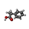  Mass: 150.174 Da / Num. of mol.: 1 / Source method: obtained synthetically / Formula: C9H10O2 Mass: 150.174 Da / Num. of mol.: 1 / Source method: obtained synthetically / Formula: C9H10O2 |
| #5: Water | ChemComp-HOH /  Mass: 18.015 Da / Num. of mol.: 360 / Source method: isolated from a natural source / Formula: H2O Mass: 18.015 Da / Num. of mol.: 360 / Source method: isolated from a natural source / Formula: H2O |
-Experimental details
-Experiment
| Experiment | Method: X-RAY DIFFRACTION / Number of used crystals: 1 |
|---|
- Sample preparation
| Crystal | Density Matthews: 2.11 Å3/Da / Density % sol: 41.1 % | ||||||||||||||||||||||||||||||
|---|---|---|---|---|---|---|---|---|---|---|---|---|---|---|---|---|---|---|---|---|---|---|---|---|---|---|---|---|---|---|---|
| Crystal grow | Temperature: 277 K / Method: vapor diffusion, hanging drop / pH: 7.2 Details: MOPS BUFFER, PEG MME 2K, PPA, pH 7.20, VAPOR DIFFUSION, HANGING DROP, temperature 277K | ||||||||||||||||||||||||||||||
| Crystal grow | *PLUS Temperature: 4 ℃ / pH: 7.2 / Method: vapor diffusion, hanging drop | ||||||||||||||||||||||||||||||
| Components of the solutions | *PLUS
|
-Data collection
| Diffraction | Mean temperature: 120 K |
|---|---|
| Diffraction source | Source: ROTATING ANODE / Type: ENRAF-NONIUS FR591 / Wavelength: 1.5418 / Wavelength: 1.5418 Å |
| Detector | Type: MAC Science DIP-2020 / Detector: IMAGE PLATE / Date: Jan 1, 2000 / Details: MIRRORS |
| Radiation | Monochromator: MIRRORS / Protocol: SINGLE WAVELENGTH / Monochromatic (M) / Laue (L): M / Scattering type: x-ray |
| Radiation wavelength | Wavelength: 1.5418 Å / Relative weight: 1 |
| Reflection | Resolution: 2.43→30 Å / Num. all: 27684 / Num. obs: 26632 / % possible obs: 96.2 % / Observed criterion σ(I): -3 / Redundancy: 2.185 % / Rsym value: 0.037 / Net I/σ(I): 21.1 |
| Reflection shell | Resolution: 2.43→2.47 Å / Mean I/σ(I) obs: 10.9 / Rsym value: 0.066 / % possible all: 95.7 |
| Reflection | *PLUS Highest resolution: 2.43 Å / Lowest resolution: 30 Å / Num. measured all: 58181 / Rmerge(I) obs: 0.037 |
| Reflection shell | *PLUS % possible obs: 95.7 % / Rmerge(I) obs: 0.066 / Mean I/σ(I) obs: 10.9 |
- Processing
| Software |
| |||||||||||||||||||||||||||||||||||||||||||||||||||||||||||||||
|---|---|---|---|---|---|---|---|---|---|---|---|---|---|---|---|---|---|---|---|---|---|---|---|---|---|---|---|---|---|---|---|---|---|---|---|---|---|---|---|---|---|---|---|---|---|---|---|---|---|---|---|---|---|---|---|---|---|---|---|---|---|---|---|---|
| Refinement | Method to determine structure: MOLECULAR REPLACEMENT Starting model: PDB ENTRY 1PNK Resolution: 2.43→30 Å / Cross valid method: THROUGHOUT / Stereochemistry target values: engh & huber
| |||||||||||||||||||||||||||||||||||||||||||||||||||||||||||||||
| Displacement parameters | Biso mean: 18.104 Å2
| |||||||||||||||||||||||||||||||||||||||||||||||||||||||||||||||
| Refine analyze | Luzzati sigma a obs: 0.21 Å | |||||||||||||||||||||||||||||||||||||||||||||||||||||||||||||||
| Refinement step | Cycle: LAST / Resolution: 2.43→30 Å
| |||||||||||||||||||||||||||||||||||||||||||||||||||||||||||||||
| Refine LS restraints |
| |||||||||||||||||||||||||||||||||||||||||||||||||||||||||||||||
| LS refinement shell | Highest resolution: 2.43 Å / Rfactor Rfree: 0.269 / Rfactor Rwork: 0.173 / Total num. of bins used: 20 | |||||||||||||||||||||||||||||||||||||||||||||||||||||||||||||||
| Software | *PLUS Version: 5 / Classification: refinement | |||||||||||||||||||||||||||||||||||||||||||||||||||||||||||||||
| Refine LS restraints | *PLUS
|
