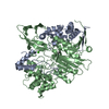
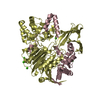
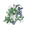
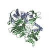
Entry Database : PDB / ID : 6nvwTitle Crystal structure of penicillin G acylase from Bacillus megaterium (Penicillin G acylase) x 2 Keywords / / Function / homology Function Domain/homology Component
/ / / / / / / / / / / / / / / / / / / / / / / / / / / / / Biological species Bacillus megaterium (bacteria)Method / / / / Resolution : 2.203 Å Authors Blankenfeldt, W. Funding support Organization Grant number Country German Research Foundation SPP1934
Journal : Appl.Microbiol.Biotechnol. / Year : 2019Title : Crystal structures and protein engineering of three different penicillin G acylases from Gram-positive bacteria with different thermostability.Authors : Mayer, J. / Pippel, J. / Gunther, G. / Muller, C. / Lauermann, A. / Knuuti, T. / Blankenfeldt, W. / Jahn, D. / Biedendieck, R. History Deposition Feb 5, 2019 Deposition site / Processing site Revision 1.0 Jul 3, 2019 Provider / Type Revision 1.1 Sep 11, 2019 Group / Database references / Category / citation_authorItem _citation.journal_volume / _citation.page_first ... _citation.journal_volume / _citation.page_first / _citation.page_last / _citation_author.identifier_ORCID Revision 1.2 Nov 20, 2019 Group / Category / Item Revision 1.3 May 15, 2024 Group / Data collection / Database referencesCategory chem_comp_atom / chem_comp_bond ... chem_comp_atom / chem_comp_bond / database_2 / pdbx_unobs_or_zero_occ_atoms Item / _database_2.pdbx_database_accession
Show all Show less
 Movie
Movie Controller

 Yorodumi
 Open data
 Basic information
 Components
 Keywords
 Function and homology information
 Bacillus megaterium (bacteria)
 X-RAY DIFFRACTION /
  Authors
 Germany, 1items
  Citation
 Structure visualization
 Downloads & links
 Other downloads


 PDBj
PDBj


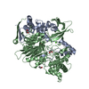
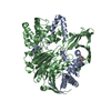
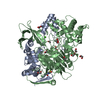
 Assembly
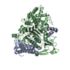

 Mass: 40.078 Da / Num. of mol.: 2 / Source method: obtained synthetically / Formula: Ca
 Mass: 18.015 Da / Num. of mol.: 256 / Source method: isolated from a natural source / Formula: H2O
 Sample preparation
 / Beamline: X06DA / Wavelength: 1.0003 Å
 Processing