 Movie
Movie Controller

[English] 日本語
 Yorodumi
- PDB-3me6: Crystal structure of cytochrome 2B4 in complex with the anti-plat... -
+ Open data

- Basic information
| Entry | Database: PDB / ID: 3me6 | ||||||
|---|---|---|---|---|---|---|---|
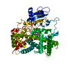
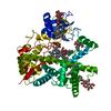
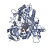
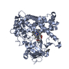
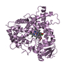
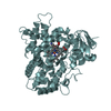
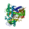
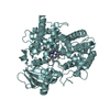
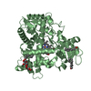
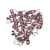
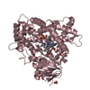
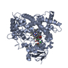
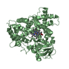
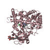
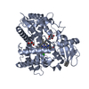
| Title | Crystal structure of cytochrome 2B4 in complex with the anti-platelet drug clopidogrel | ||||||
 Components Components | Cytochrome P450 2B4 | ||||||
 Keywords Keywords | OXIDOREDUCTASE / P450 / cytochrome P450 2B4 / monooxygenase / membrane protein / CYP 2B4 / CYP LM2 | ||||||
| Function / homology |  Function and homology information Function and homology informationarachidonate epoxygenase activity / epoxygenase P450 pathway / oxidoreductase activity, acting on paired donors, with incorporation or reduction of molecular oxygen, reduced flavin or flavoprotein as one donor, and incorporation of one atom of oxygen / unspecific monooxygenase / xenobiotic metabolic process / iron ion binding / heme binding / endoplasmic reticulum membrane Similarity search - Function | ||||||
| Biological species |  | ||||||
| Method |  X-RAY DIFFRACTION / SYNCHROTRON / MOLECULAR REPLACEMENT / molecular replacement / Resolution: 3.1 Å X-RAY DIFFRACTION / SYNCHROTRON / MOLECULAR REPLACEMENT / molecular replacement / Resolution: 3.1 Å | ||||||
 Authors Authors | Gay, S.C. / Roberts, A.G. / Maekawa, K. / Talakad, J.C. / Hong, W.X. / Zhang, Q. / Stout, C.D. / Halpert, J.R. | ||||||
 Citation Citation | Journal: Biochemistry / Year: 2010 Title: Structures of cytochrome P450 2B4 complexed with the antiplatelet drugs ticlopidine and clopidogrel. Authors: Gay, S.C. / Roberts, A.G. / Maekawa, K. / Talakad, J.C. / Hong, W.X. / Zhang, Q. / Stout, C.D. / Halpert, J.R. #1: Journal: J.Biol.Chem. / Year: 2006Title: Structure of microsomal cytochrome P450 2B4 complexed with the antifungal drug bifonazole: insight into P450 conformational plasticity and membrane interaction. Authors: Zhao, Y. / White, M.A. / Muralidhara, B.K. / Sun, L. / Halpert, J.R. / Stout, C.D. #2: Journal: J.Biol.Chem. / Year: 2004Title: Structure of mammalian cytochrome P450 2B4 complexed with 4-(4-chlorophenyl)imidazole at 1.9 {angstrom} resolution: Insight into the range of P450 conformations and coordination of redox partner binding. Authors: Scott, E.E. / White, M.A. / He, Y.A. / Johnson, E.F. / Stout, C.D. / Halpert, J.R. #3: Journal: Proc.Natl.Acad.Sci.USA / Year: 2003Title: An open conformation of mammalian cytochrome P450 2B4 at 1.6 A resolution. Authors: Scott, E.E. / He, Y.A. / Wester, M.R. / White, M.A. / Chin, C.C. / Halpert, J.R. / Johnson, E.F. / Stout, C.D. #4: Journal: Biochemistry / Year: 2007Title: Structural and thermodynamic consequences of 1-(4-chlorophenyl)imidazole binding to cytochrome P450 2B4. Authors: Zhao, Y. / Sun, L. / Muralidhara, B.K. / White, M.A. / Stout, C.D. / Halpert, J.R. #5: Journal: Biochemistry / Year: 2009Title: Crystal Structures of Cytochrome P450 2B4 in Complex with the Inhibitor 1-Biphenyl-4-methyl-1H-imidazole: Ligand-Induced Structural Response through Alpha-Helical Repositioning Authors: Gay, S.C. / Sun, L. / Maekawa, K. / Halpert, J.R. / Stout, C.D. | ||||||
| History |
|
- Structure visualization
| Structure viewer | Molecule: MolmilJmol/JSmol |
|---|
- Downloads & links
-Download
| PDBx/mmCIF format | 3me6.cif.gz | 381.2 KB | Display | PDBx/mmCIF format |
|---|---|---|---|---|
| PDB format | pdb3me6.ent.gz | 312.1 KB | Display | PDB format |
| PDBx/mmJSON format | 3me6.json.gz | Tree view | PDBx/mmJSON format | |
| Others |  Other downloads Other downloads |
-Validation report
| Summary document | 3me6_validation.pdf.gz | 2.8 MB | Display | wwPDB validaton report |
|---|---|---|---|---|
| Full document | 3me6_full_validation.pdf.gz | 2.9 MB | Display | |
| Data in XML | 3me6_validation.xml.gz | 75.4 KB | Display | |
| Data in CIF | 3me6_validation.cif.gz | 97.7 KB | Display | |
| Arichive directory | https://data.pdbj.org/pub/pdb/validation_reports/me/3me6ftp://data.pdbj.org/pub/pdb/validation_reports/me/3me6 | HTTPS FTP |
-Related structure data
| Related structure data | 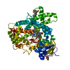 3kw4C 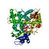 1suoS S: Starting model for refinement C: citing same article ( |
|---|---|
| Similar structure data |


-Links
 PDBj
PDBj


- Assembly
| Deposited unit | 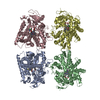
| ||||||||||||||||||||||||||||||
|---|---|---|---|---|---|---|---|---|---|---|---|---|---|---|---|---|---|---|---|---|---|---|---|---|---|---|---|---|---|---|---|
| 1 | 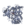
| ||||||||||||||||||||||||||||||
| 2 | 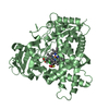
| ||||||||||||||||||||||||||||||
| 3 | 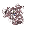
| ||||||||||||||||||||||||||||||
| 4 | 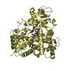
| ||||||||||||||||||||||||||||||
| Unit cell |
| ||||||||||||||||||||||||||||||
| Noncrystallographic symmetry (NCS) | NCS domain:
NCS domain segments: Component-ID: 1 / Ens-ID: 1 / Beg auth comp-ID: GLY / Beg label comp-ID: GLY / End auth comp-ID: HIS / End label comp-ID: HIS / Refine code: 6 / Auth seq-ID: 28 - 492 / Label seq-ID: 9 - 473
|
-Components
| #1: Protein | Mass: 54169.082 Da / Num. of mol.: 4 Mutation: E2A, G22K, H23K, P24T, K25S, A26S, H27K, R29K, H226Y Source method: isolated from a genetically manipulated source Details: Deleted residues 3-21, E2A, G22K, H23K, P24T, K25S, A26S, H27K, R29K, 4x Cterm His tag Source: (gene. exp.)  #2: Chemical | ChemComp-HEM /   Mass: 616.487 Da / Num. of mol.: 4 / Source method: obtained synthetically / Formula: C34H32FeN4O4 Mass: 616.487 Da / Num. of mol.: 4 / Source method: obtained synthetically / Formula: C34H32FeN4O4#3: Chemical | ChemComp-CGE /   Mass: 321.822 Da / Num. of mol.: 4 / Source method: obtained synthetically / Formula: C16H16ClNO2S Mass: 321.822 Da / Num. of mol.: 4 / Source method: obtained synthetically / Formula: C16H16ClNO2S#4: Water | ChemComp-HOH / |  Mass: 18.015 Da / Num. of mol.: 132 / Source method: isolated from a natural source / Formula: H2O Mass: 18.015 Da / Num. of mol.: 132 / Source method: isolated from a natural source / Formula: H2OSequence details | AUTHORS INDICATE THAT THERE IS AN ERROR IN THE SEQUENCE DATABASE REFERENCE AT RESIDUE 221 | |
|---|
-Experimental details
-Experiment
| Experiment | Method: X-RAY DIFFRACTION / Number of used crystals: 1 |
|---|
- Sample preparation
| Crystal | Density Matthews: 4.2 Å3/Da / Density % sol: 70.71 % |
|---|---|
| Crystal grow | Temperature: 291 K / Method: vapor diffusion, sitting drop / pH: 4.2 Details: 20% (w/v) PEG 8000, 0.2 M NaCl and 0.1 M phosphate-citrate, pH 4.2, vapor diffusion, sitting drop, temperature 291K |
-Data collection
| Diffraction | Mean temperature: 100 K |
|---|---|
| Diffraction source | Source: SYNCHROTRON / Site: SSRL  / Beamline: BL11-1 / Wavelength: 0.98 Å / Beamline: BL11-1 / Wavelength: 0.98 Å |
| Detector | Type: MARMOSAIC 325 mm CCD / Detector: CCD / Date: Nov 7, 2009 |
| Radiation | Monochromator: Si(111) / Protocol: SINGLE WAVELENGTH / Monochromatic (M) / Laue (L): M / Scattering type: x-ray |
| Radiation wavelength | Wavelength: 0.98 Å / Relative weight: 1 |
| Reflection | Resolution: 3.1→76.755 Å / Num. all: 62529 / Num. obs: 62529 / % possible obs: 97.7 % / Observed criterion σ(F): 0 / Observed criterion σ(I): 0 / Redundancy: 4 % / Rmerge(I) obs: 0.13 / Rsym value: 0.13 / Net I/σ(I): 4.7 |
| Reflection shell | Resolution: 3.1→3.18 Å / Redundancy: 3.9 % / Rmerge(I) obs: 0.432 / Mean I/σ(I) obs: 3.5 / Num. unique all: 9259 / Rsym value: 0.432 / % possible all: 98.6 |
-Phasing
| Phasing | Method: molecular replacement | |||||||||
|---|---|---|---|---|---|---|---|---|---|---|
| Phasing MR | Rfactor: 33.25 / Model details: Phaser MODE: MR_AUTO
|
- Processing
| Software |
| |||||||||||||||||||||||||||||||||||||||||||||||||||||||||||||||||
|---|---|---|---|---|---|---|---|---|---|---|---|---|---|---|---|---|---|---|---|---|---|---|---|---|---|---|---|---|---|---|---|---|---|---|---|---|---|---|---|---|---|---|---|---|---|---|---|---|---|---|---|---|---|---|---|---|---|---|---|---|---|---|---|---|---|---|
| Refinement | Method to determine structure: MOLECULAR REPLACEMENT Starting model: 1SUO Resolution: 3.1→76.75 Å / Cor.coef. Fo:Fc: 0.933 / Cor.coef. Fo:Fc free: 0.885 / WRfactor Rfree: 0.237 / WRfactor Rwork: 0.181 / Occupancy max: 1 / Occupancy min: 0.5 / FOM work R set: 0.857 / SU B: 14.228 / SU ML: 0.258 / SU Rfree: 0.378 / Cross valid method: THROUGHOUT / σ(F): 0 / σ(I): 0 / ESU R Free: 0.378 / Stereochemistry target values: MAXIMUM LIKELIHOOD Details: HYDROGENS HAVE BEEN ADDED IN THE RIDING POSITIONS. U VALUES: REFINED INDIVIDUALLY
| |||||||||||||||||||||||||||||||||||||||||||||||||||||||||||||||||
| Solvent computation | Ion probe radii: 0.8 Å / Shrinkage radii: 0.8 Å / VDW probe radii: 1.4 Å / Solvent model: MASK | |||||||||||||||||||||||||||||||||||||||||||||||||||||||||||||||||
| Displacement parameters | Biso max: 111.96 Å2 / Biso mean: 42.197 Å2 / Biso min: 2 Å2
| |||||||||||||||||||||||||||||||||||||||||||||||||||||||||||||||||
| Refinement step | Cycle: LAST / Resolution: 3.1→76.75 Å
| |||||||||||||||||||||||||||||||||||||||||||||||||||||||||||||||||
| Refine LS restraints |
| |||||||||||||||||||||||||||||||||||||||||||||||||||||||||||||||||
| Refine LS restraints NCS | Ens-ID: 1 / Number: 3718 / Refine-ID: X-RAY DIFFRACTION
| |||||||||||||||||||||||||||||||||||||||||||||||||||||||||||||||||
| LS refinement shell | Resolution: 3.1→3.181 Å / Total num. of bins used: 20
|
