 Movie
Movie Controller


+ Open data

- Basic information
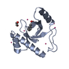
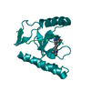
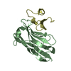
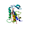
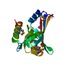
| Entry | Database: PDB / ID: 3lyi | ||||||
|---|---|---|---|---|---|---|---|
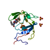
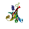
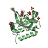
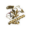
| Title | PWWP Domain of Human Bromodomain-Containing Protein 1 | ||||||
 Components Components | Bromodomain-containing protein 1 | ||||||
 Keywords Keywords | TRANSCRIPTION / histone H3 acetylation / Structural Genomics Consortium / SGC / Bromodomain / Chromatin regulator | ||||||
| Function / homology |  Function and homology information Function and homology informationunmodified histone reader activity / histone H3-K14 acetyltransferase complex / MOZ/MORF histone acetyltransferase complex / regulation of hemopoiesis / regulation of developmental process / erythrocyte maturation / response to immobilization stress / response to electrical stimulus / histone reader activity / Regulation of TP53 Activity through Acetylation ...unmodified histone reader activity / histone H3-K14 acetyltransferase complex / MOZ/MORF histone acetyltransferase complex / regulation of hemopoiesis / regulation of developmental process / erythrocyte maturation / response to immobilization stress / response to electrical stimulus / histone reader activity / Regulation of TP53 Activity through Acetylation / positive regulation of erythrocyte differentiation / HATs acetylate histones / perikaryon / nuclear speck / chromatin remodeling / regulation of transcription by RNA polymerase II / regulation of DNA-templated transcription / dendrite / zinc ion binding / nucleus Similarity search - Function | ||||||
| Biological species |  Homo sapiens (human) Homo sapiens (human) | ||||||
| Method |  X-RAY DIFFRACTION / SYNCHROTRON / MOLECULAR REPLACEMENT / Resolution: 2.1 Å X-RAY DIFFRACTION / SYNCHROTRON / MOLECULAR REPLACEMENT / Resolution: 2.1 Å | ||||||
 Authors Authors | Lam, R. / Zeng, H. / Ni, S. / Bountra, C. / Weigelt, J. / Arrowsmith, C.H. / Edwards, A.M. / Bochkarev, A. / Min, J. / Wu, H. / Structural Genomics Consortium (SGC) | ||||||
 Citation Citation | Journal: Plos One / Year: 2011 Title: Structural and histone binding ability characterizations of human PWWP domains. Authors: Wu, H. / Zeng, H. / Lam, R. / Tempel, W. / Amaya, M.F. / Xu, C. / Dombrovski, L. / Qiu, W. / Wang, Y. / Min, J. | ||||||
| History |
|
- Structure visualization
| Structure viewer | Molecule: MolmilJmol/JSmol |
|---|
- Downloads & links
-Download
| PDBx/mmCIF format | 3lyi.cif.gz | 108.4 KB | Display | PDBx/mmCIF format |
|---|---|---|---|---|
| PDB format | pdb3lyi.ent.gz | 83.3 KB | Display | PDB format |
| PDBx/mmJSON format | 3lyi.json.gz | Tree view | PDBx/mmJSON format | |
| Others |  Other downloads Other downloads |
-Validation report
| Arichive directory | https://data.pdbj.org/pub/pdb/validation_reports/ly/3lyiftp://data.pdbj.org/pub/pdb/validation_reports/ly/3lyi | HTTPS FTP |
|---|
-Related structure data
| Related structure data |  3eaeC 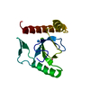 3l42SC 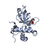 3llrC 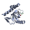 3mo8C 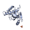 3pfsC 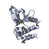 3pmiC  3qbyC  3qj6C  3qkjC S: Starting model for refinement C: citing same article ( |
|---|---|
| Similar structure data |

-Links
 PDBj
PDBj


- Assembly
| Deposited unit | 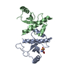
| ||||||||
|---|---|---|---|---|---|---|---|---|---|
| 1 | 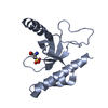
| ||||||||
| 2 | 
| ||||||||
| Unit cell |
|
-Components
| #1: Protein | Mass: 14308.836 Da / Num. of mol.: 2 / Fragment: PWWP Domain, residues 925-1049 Source method: isolated from a genetically manipulated source Source: (gene. exp.) Homo sapiens (human) / Gene: BRD1, BRL, BRPF2 / Plasmid: pET28-MHL / Production host:  #2: Chemical | ChemComp-OCS / |   Type: L-peptide linking / Mass: 169.156 Da / Num. of mol.: 1 / Source method: obtained synthetically / Formula: C3H7NO5S Type: L-peptide linking / Mass: 169.156 Da / Num. of mol.: 1 / Source method: obtained synthetically / Formula: C3H7NO5S#3: Water | ChemComp-HOH / |  Mass: 18.015 Da / Num. of mol.: 78 / Source method: isolated from a natural source / Formula: H2O Mass: 18.015 Da / Num. of mol.: 78 / Source method: isolated from a natural source / Formula: H2O |
|---|
-Experimental details
-Experiment
| Experiment | Method: X-RAY DIFFRACTION / Number of used crystals: 1 |
|---|
- Sample preparation
| Crystal | Density Matthews: 2.37 Å3/Da / Density % sol: 48.09 % |
|---|---|
| Crystal grow | Temperature: 293 K / Method: vapor diffusion, sitting drop / pH: 7.5 Details: 30% PEG2000-MME, 0.15M KBr, pH 7.5, VAPOR DIFFUSION, SITTING DROP, temperature 293K |
-Data collection
| Diffraction | Mean temperature: 100 K | |||||||||||||||||||||||||||||||||||||||||||||||||||||||||||||||||||||||||||||
|---|---|---|---|---|---|---|---|---|---|---|---|---|---|---|---|---|---|---|---|---|---|---|---|---|---|---|---|---|---|---|---|---|---|---|---|---|---|---|---|---|---|---|---|---|---|---|---|---|---|---|---|---|---|---|---|---|---|---|---|---|---|---|---|---|---|---|---|---|---|---|---|---|---|---|---|---|---|---|
| Diffraction source | Source: SYNCHROTRON / Site: CLSI  / Beamline: 08ID-1 / Wavelength: 0.97949 Å / Beamline: 08ID-1 / Wavelength: 0.97949 Å | |||||||||||||||||||||||||||||||||||||||||||||||||||||||||||||||||||||||||||||
| Detector | Type: MARMOSAIC 225 mm CCD / Detector: CCD / Date: Jan 24, 2010 | |||||||||||||||||||||||||||||||||||||||||||||||||||||||||||||||||||||||||||||
| Radiation | Monochromator: double crystal monochromator / Protocol: SINGLE WAVELENGTH / Monochromatic (M) / Laue (L): M / Scattering type: x-ray | |||||||||||||||||||||||||||||||||||||||||||||||||||||||||||||||||||||||||||||
| Radiation wavelength | Wavelength: 0.97949 Å / Relative weight: 1 | |||||||||||||||||||||||||||||||||||||||||||||||||||||||||||||||||||||||||||||
| Reflection | Resolution: 2.1→50 Å / Num. obs: 16475 / % possible obs: 99 % / Redundancy: 6.7 % / Rmerge(I) obs: 0.074 / Χ2: 1.007 / Net I/σ(I): 11.5 | |||||||||||||||||||||||||||||||||||||||||||||||||||||||||||||||||||||||||||||
| Reflection shell |
|
-Phasing
| Phasing MR |
|
|---|
- Processing
| Software |
| |||||||||||||||||||||||||||||||||||||||||||||||||||||||||||||||||||||||||||
|---|---|---|---|---|---|---|---|---|---|---|---|---|---|---|---|---|---|---|---|---|---|---|---|---|---|---|---|---|---|---|---|---|---|---|---|---|---|---|---|---|---|---|---|---|---|---|---|---|---|---|---|---|---|---|---|---|---|---|---|---|---|---|---|---|---|---|---|---|---|---|---|---|---|---|---|---|
| Refinement | Method to determine structure: MOLECULAR REPLACEMENT Starting model: 3L42 Resolution: 2.1→37.66 Å / Cor.coef. Fo:Fc: 0.936 / Cor.coef. Fo:Fc free: 0.91 / Occupancy max: 1 / Occupancy min: 1 / SU B: 13.125 / SU ML: 0.159 / Cross valid method: THROUGHOUT / σ(F): 0 / ESU R: 0.242 / ESU R Free: 0.205 / Stereochemistry target values: MAXIMUM LIKELIHOOD Details: HYDROGENS HAVE BEEN ADDED IN THE RIDING POSITIONS; U VALUES: WITH TLS ADDED. unknown ligand was not modeled.
| |||||||||||||||||||||||||||||||||||||||||||||||||||||||||||||||||||||||||||
| Solvent computation | Ion probe radii: 0.8 Å / Shrinkage radii: 0.8 Å / VDW probe radii: 1.4 Å / Solvent model: BABINET MODEL WITH MASK | |||||||||||||||||||||||||||||||||||||||||||||||||||||||||||||||||||||||||||
| Displacement parameters | Biso max: 124.8 Å2 / Biso mean: 59.069 Å2 / Biso min: 24.65 Å2
| |||||||||||||||||||||||||||||||||||||||||||||||||||||||||||||||||||||||||||
| Refinement step | Cycle: LAST / Resolution: 2.1→37.66 Å
| |||||||||||||||||||||||||||||||||||||||||||||||||||||||||||||||||||||||||||
| Refine LS restraints |
| |||||||||||||||||||||||||||||||||||||||||||||||||||||||||||||||||||||||||||
| LS refinement shell | Resolution: 2.1→2.151 Å / Total num. of bins used: 20
| |||||||||||||||||||||||||||||||||||||||||||||||||||||||||||||||||||||||||||
| Refinement TLS params. | Method: refined / Refine-ID: X-RAY DIFFRACTION
| |||||||||||||||||||||||||||||||||||||||||||||||||||||||||||||||||||||||||||
| Refinement TLS group |
|
