Resolution: 1.3→40 Å / Num. obs: 31403 / % possible obs: 99.9 % / Redundancy: 14.5 % / Rmerge(I) obs: 0.056 / Χ2: 1.614 / Net I/σ(I): 12.2
Reflection shell
Resolution (Å)
Redundancy (%)
Rmerge(I) obs
Num. unique all
Χ2
Diffraction-ID
% possible all
1.3-1.32
13.8
0.465
1544
1.468
1,2
100
1.32-1.35
14.2
0.387
1535
1.465
1,2
100
1.35-1.37
14.3
0.346
1522
1.473
1,2
100
1.37-1.4
14.3
0.294
1544
1.375
1,2
100
1.4-1.43
14.3
0.253
1548
1.353
1,2
100
1.43-1.46
14.4
0.218
1521
1.302
1,2
100
1.46-1.5
14.3
0.169
1544
1.272
1,2
100
1.5-1.54
14.4
0.143
1549
1.285
1,2
100
1.54-1.59
14.4
0.124
1544
1.217
1,2
100
1.59-1.64
14.6
0.103
1542
1.18
1,2
100
1.64-1.7
14.5
0.092
1562
1.2
1,2
100
1.7-1.76
14.6
0.079
1548
1.199
1,2
100
1.76-1.84
14.7
0.068
1580
1.252
1,2
100
1.84-1.94
14.9
0.058
1545
1.365
1,2
100
1.94-2.06
15
0.057
1569
1.651
1,2
100
2.06-2.22
15
0.059
1600
2.209
1,2
100
2.22-2.45
15
0.061
1583
2.633
1,2
100
2.45-2.8
14.8
0.052
1614
2.424
1,2
100
2.8-3.53
14.3
0.042
1649
2.33
1,2
99.9
3.53-40
13.4
0.039
1760
2.384
1,2
98.5
-
Phasing
Phasing
Method: sad
-
Processing
Software
Name
Version
Classification
NB
DENZO
datareduction
SCALEPACK
datascaling
SHELX
phasing
REFMAC
5.5.0102
refinement
PDB_EXTRACT
3.005
dataextraction
HKL-2000
datascaling
Refinement
Method to determine structure: SAD / Resolution: 1.3→30 Å / Cor.coef. Fo:Fc: 0.947 / Cor.coef. Fo:Fc free: 0.92 / WRfactor Rfree: 0.234 / WRfactor Rwork: 0.211 / SU B: 0.726 / SU ML: 0.032 / Cross valid method: THROUGHOUT / σ(F): 0 / ESU R: 0.06 / ESU R Free: 0.061 / Stereochemistry target values: MAXIMUM LIKELIHOOD Details: HYDROGENS HAVE BEEN ADDED IN THE RIDING POSITIONS U VALUES: REFINED INDIVIDUALLY The structure was solved using the selenomethionene derivative crystallized in space group I222 and a copper ...Details: HYDROGENS HAVE BEEN ADDED IN THE RIDING POSITIONS U VALUES: REFINED INDIVIDUALLY The structure was solved using the selenomethionene derivative crystallized in space group I222 and a copper rotating anode. The programs ARP/WARP, COOT and MOLPROBITY were also used during model refinement.
Rfactor
Num. reflection
% reflection
Selection details
Rfree
0.233
1565
5.034 %
RANDOM
Rwork
0.209
-
-
-
obs
0.21
31089
99.926 %
-
Solvent computation
Ion probe radii: 0.8 Å / Shrinkage radii: 0.8 Å / VDW probe radii: 1.4 Å / Solvent model: MASK BULK SOLVENT
Displacement parameters
Biso mean: 10.096 Å2
Baniso -1
Baniso -2
Baniso -3
1-
-0.007 Å2
0 Å2
0 Å2
2-
-
-0.007 Å2
0 Å2
3-
-
-
0.015 Å2
Refinement step
Cycle: LAST / Resolution: 1.3→30 Å
Protein
Nucleic acid
Ligand
Solvent
Total
Num. atoms
983
0
3
108
1094
Refine LS restraints
Refine-ID
Type
Dev ideal
Dev ideal target
Number
X-RAY DIFFRACTION
r_bond_refined_d
0.016
0.022
1099
X-RAY DIFFRACTION
r_bond_other_d
0.001
0.02
752
X-RAY DIFFRACTION
r_angle_refined_deg
1.632
1.977
1519
X-RAY DIFFRACTION
r_angle_other_deg
0.968
3
1864
X-RAY DIFFRACTION
r_dihedral_angle_1_deg
5.307
5
153
X-RAY DIFFRACTION
r_dihedral_angle_2_deg
36.245
23.636
44
X-RAY DIFFRACTION
r_dihedral_angle_3_deg
13.53
15
189
X-RAY DIFFRACTION
r_dihedral_angle_4_deg
9.874
15
6
X-RAY DIFFRACTION
r_chiral_restr
0.103
0.2
172
X-RAY DIFFRACTION
r_gen_planes_refined
0.009
0.021
1226
X-RAY DIFFRACTION
r_gen_planes_other
0.001
0.02
213
X-RAY DIFFRACTION
r_mcbond_it
1.096
1.5
680
X-RAY DIFFRACTION
r_mcbond_other
0.308
1.5
263
X-RAY DIFFRACTION
r_mcangle_it
1.855
2
1115
X-RAY DIFFRACTION
r_scbond_it
2.443
3
419
X-RAY DIFFRACTION
r_scangle_it
3.455
4.5
390
LS refinement shell
Refine-ID: X-RAY DIFFRACTION / Total num. of bins used: 20
Resolution (Å)
Rfactor Rfree
Num. reflection Rfree
Rfactor Rwork
Num. reflection Rwork
Num. reflection all
% reflection obs (%)
1.3-1.334
0.248
119
0.255
2131
2250
100
1.334-1.37
0.269
107
0.232
2071
2178
100
1.37-1.41
0.238
101
0.231
2024
2125
100
1.41-1.453
0.218
112
0.214
1964
2076
100
1.453-1.501
0.245
117
0.201
1918
2035
100
1.501-1.553
0.216
87
0.179
1850
1937
100
1.553-1.612
0.229
78
0.195
1823
1901
100
1.612-1.677
0.193
93
0.18
1716
1809
100
1.677-1.752
0.239
87
0.191
1664
1751
100
1.752-1.837
0.231
85
0.192
1616
1701
100
1.837-1.936
0.214
86
0.195
1506
1592
100
1.936-2.053
0.197
82
0.197
1435
1517
100
2.053-2.194
0.195
69
0.197
1374
1443
100
2.194-2.368
0.19
72
0.206
1270
1342
100
2.368-2.593
0.22
54
0.195
1204
1258
100
2.593-2.896
0.243
62
0.217
1065
1127
100
2.896-3.339
0.19
52
0.217
974
1026
100
3.339-4.077
0.264
55
0.203
837
893
99.888
4.077-5.713
0.287
23
0.21
680
704
99.858
5.713-30
0.429
24
0.344
402
447
95.302
+
About Yorodumi
-
News
-
Feb 9, 2022. New format data for meta-information of EMDB entries
New format data for meta-information of EMDB entries
Version 3 of the EMDB header file is now the official format.
The previous official version 1.9 will be removed from the archive.
In the structure databanks used in Yorodumi, some data are registered as the other names, "COVID-19 virus" and "2019-nCoV". Here are the details of the virus and the list of structure data.
Jan 31, 2019. EMDB accession codes are about to change! (news from PDBe EMDB page)
EMDB accession codes are about to change! (news from PDBe EMDB page)
The allocation of 4 digits for EMDB accession codes will soon come to an end. Whilst these codes will remain in use, new EMDB accession codes will include an additional digit and will expand incrementally as the available range of codes is exhausted. The current 4-digit format prefixed with “EMD-” (i.e. EMD-XXXX) will advance to a 5-digit format (i.e. EMD-XXXXX), and so on. It is currently estimated that the 4-digit codes will be depleted around Spring 2019, at which point the 5-digit format will come into force.
The EM Navigator/Yorodumi systems omit the EMD- prefix.
Related info.:Q: What is EMD? / ID/Accession-code notation in Yorodumi/EM Navigator
Yorodumi is a browser for structure data from EMDB, PDB, SASBDB, etc.
This page is also the successor to EM Navigator detail page, and also detail information page/front-end page for Omokage search.
The word "yorodu" (or yorozu) is an old Japanese word meaning "ten thousand". "mi" (miru) is to see.
Related info.:EMDB / PDB / SASBDB / Comparison of 3 databanks / Yorodumi Search / Aug 31, 2016. New EM Navigator & Yorodumi / Yorodumi Papers / Jmol/JSmol / Function and homology information / Changes in new EM Navigator and Yorodumi
 Movie
Movie Controller

 Yorodumi
 Open data
 Basic information
 Components
 Keywords
 Function and homology information
 Homo sapiens (human)
 X-RAY DIFFRACTION /
  Authors
 Citation
 Structure visualization
 Downloads & links
 Other downloads


 PDBj
PDBj


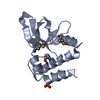
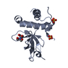
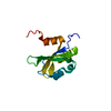
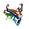
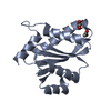
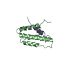
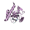
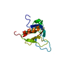
 Assembly


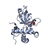
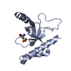
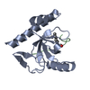
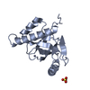
 Num. of mol.: 3 / Source method: obtained synthetically
 Mass: 18.015 Da / Num. of mol.: 108 / Source method: isolated from a natural source / Formula: H2O
 Sample preparation
 Processing