 Movie
Movie Controller

[English] 日本語
 Yorodumi
- PDB-3lif: Crystal Structure of the extracellular domain of the putative his... -
+ Open data

- Basic information
| Entry | Database: PDB / ID: 3lif | ||||||
|---|---|---|---|---|---|---|---|
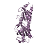
| Title | Crystal Structure of the extracellular domain of the putative histidine kinase rpHK1S-Z16 | ||||||
 Components Components | Putative diguanylate cyclase (GGDEF) with PAS/PAC domain | ||||||
 Keywords Keywords | SIGNALING PROTEIN / PDC fold | ||||||
| Function / homology |  Function and homology information Function and homology informationnegative regulation of bacterial-type flagellum-dependent cell motility / diguanylate cyclase / diguanylate cyclase activity / cell adhesion involved in single-species biofilm formation / plasma membrane Similarity search - Function | ||||||
| Biological species |  Rhodopseudomonas palustris (phototrophic) Rhodopseudomonas palustris (phototrophic) | ||||||
| Method |  X-RAY DIFFRACTION / SYNCHROTRON / MAD / Resolution: 2.7 Å X-RAY DIFFRACTION / SYNCHROTRON / MAD / Resolution: 2.7 Å | ||||||
 Authors Authors | Zhang, Z. / Hendrickson, W.A. | ||||||
 Citation Citation | Journal: J.Mol.Biol. / Year: 2010 Title: Structural characterization of the predominant family of histidine kinase sensor domains. Authors: Zhang, Z. / Hendrickson, W.A. | ||||||
| History |
|
- Structure visualization
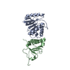
| Structure viewer | Molecule: MolmilJmol/JSmol |
|---|
- Downloads & links
-Download
| PDBx/mmCIF format | 3lif.cif.gz | 111.9 KB | Display | PDBx/mmCIF format |
|---|---|---|---|---|
| PDB format | pdb3lif.ent.gz | 87.2 KB | Display | PDB format |
| PDBx/mmJSON format | 3lif.json.gz | Tree view | PDBx/mmJSON format | |
| Others |  Other downloads Other downloads |
-Validation report
| Arichive directory | https://data.pdbj.org/pub/pdb/validation_reports/li/3lifftp://data.pdbj.org/pub/pdb/validation_reports/li/3lif | HTTPS FTP |
|---|
-Related structure data
| Related structure data |  3li8C  3li9C  3liaC 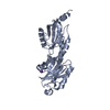 3libC  3licC 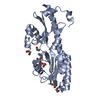 3lidC 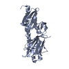 3lieC C: citing same article ( |
|---|---|
| Similar structure data |


-Links
 PDBj
PDBj


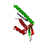
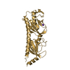
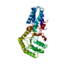
- Assembly
| Deposited unit | 
| ||||||||
|---|---|---|---|---|---|---|---|---|---|
| 1 | 
| ||||||||
| 2 | 
| ||||||||
| Unit cell |
|
-Components
| #1: Protein | Mass: 28769.756 Da / Num. of mol.: 2 / Fragment: extracellular domain (UNP residues 40-293) Source method: isolated from a genetically manipulated source Source: (gene. exp.) Rhodopseudomonas palustris (phototrophic)Gene: RPA3616 / Plasmid: pSMT3 / Production host: #2: Chemical |   Mass: 118.174 Da / Num. of mol.: 2 / Source method: obtained synthetically / Formula: C6H14O2 / Comment: precipitant*YM Mass: 118.174 Da / Num. of mol.: 2 / Source method: obtained synthetically / Formula: C6H14O2 / Comment: precipitant*YM#3: Chemical |   Mass: 192.124 Da / Num. of mol.: 2 / Source method: obtained synthetically / Formula: C6H8O7 Mass: 192.124 Da / Num. of mol.: 2 / Source method: obtained synthetically / Formula: C6H8O7#4: Water | ChemComp-HOH / |  Mass: 18.015 Da / Num. of mol.: 93 / Source method: isolated from a natural source / Formula: H2O Mass: 18.015 Da / Num. of mol.: 93 / Source method: isolated from a natural source / Formula: H2OHas protein modification | Y | |
|---|
-Experimental details
-Experiment
| Experiment | Method: X-RAY DIFFRACTION / Number of used crystals: 1 |
|---|
- Sample preparation
| Crystal | Density Matthews: 4.57 Å3/Da / Density % sol: 73.08 % |
|---|---|
| Crystal grow | Temperature: 293 K / Method: vapor diffusion / pH: 5.6 Details: 0.2M NH4Ac, 0.1M citrate pH5.6, 30% MPD, vapor diffusion, temperature 293K |
-Data collection
| Diffraction | Mean temperature: 100 K | |||||||||||||||||||||||||||||||||||||||||||||||||||||||||||||||||||||||||||||
|---|---|---|---|---|---|---|---|---|---|---|---|---|---|---|---|---|---|---|---|---|---|---|---|---|---|---|---|---|---|---|---|---|---|---|---|---|---|---|---|---|---|---|---|---|---|---|---|---|---|---|---|---|---|---|---|---|---|---|---|---|---|---|---|---|---|---|---|---|---|---|---|---|---|---|---|---|---|---|
| Diffraction source | Source: SYNCHROTRON / Site: NSLS  / Beamline: X4A / Wavelength: 0.97918, 0.97935, 0.96788 / Beamline: X4A / Wavelength: 0.97918, 0.97935, 0.96788 | |||||||||||||||||||||||||||||||||||||||||||||||||||||||||||||||||||||||||||||
| Detector | Detector: CCD | |||||||||||||||||||||||||||||||||||||||||||||||||||||||||||||||||||||||||||||
| Radiation | Protocol: MAD / Monochromatic (M) / Laue (L): M / Scattering type: x-ray | |||||||||||||||||||||||||||||||||||||||||||||||||||||||||||||||||||||||||||||
| Radiation wavelength |
| |||||||||||||||||||||||||||||||||||||||||||||||||||||||||||||||||||||||||||||
| Reflection | Redundancy: 12.9 % / Av σ(I) over netI: 43.17 / Number: 725323 / Rmerge(I) obs: 0.065 / Χ2: 1 / D res high: 2.7 Å / D res low: 50 Å / Num. obs: 56039 / % possible obs: 100 | |||||||||||||||||||||||||||||||||||||||||||||||||||||||||||||||||||||||||||||
| Diffraction reflection shell |
| |||||||||||||||||||||||||||||||||||||||||||||||||||||||||||||||||||||||||||||
| Reflection | Resolution: 2.7→50 Å / Num. obs: 56039 / % possible obs: 100 % / Redundancy: 12.9 % / Rmerge(I) obs: 0.065 / Χ2: 0.996 / Net I/σ(I): 12.4 | |||||||||||||||||||||||||||||||||||||||||||||||||||||||||||||||||||||||||||||
| Reflection shell |
|
- Processing
| Software |
| |||||||||||||||||||||||||||||||||||||||||||||||||||||||||||||||||
|---|---|---|---|---|---|---|---|---|---|---|---|---|---|---|---|---|---|---|---|---|---|---|---|---|---|---|---|---|---|---|---|---|---|---|---|---|---|---|---|---|---|---|---|---|---|---|---|---|---|---|---|---|---|---|---|---|---|---|---|---|---|---|---|---|---|---|
| Refinement | Method to determine structure: MAD / Resolution: 2.7→42.39 Å / Cor.coef. Fo:Fc: 0.936 / Cor.coef. Fo:Fc free: 0.893 / Occupancy max: 1 / Occupancy min: 0.5 / SU B: 9.23 / SU ML: 0.186 / Cross valid method: THROUGHOUT / σ(F): 0 / ESU R: 0.316 / ESU R Free: 0.262 / Stereochemistry target values: MAXIMUM LIKELIHOOD Details: HYDROGENS HAVE BEEN ADDED IN THE RIDING POSITIONS. U VALUES: REFINED INDIVIDUALLY
| |||||||||||||||||||||||||||||||||||||||||||||||||||||||||||||||||
| Solvent computation | Ion probe radii: 0.8 Å / Shrinkage radii: 0.8 Å / VDW probe radii: 1.4 Å / Solvent model: MASK | |||||||||||||||||||||||||||||||||||||||||||||||||||||||||||||||||
| Displacement parameters | Biso max: 103.57 Å2 / Biso mean: 41.753 Å2 / Biso min: 2 Å2 | |||||||||||||||||||||||||||||||||||||||||||||||||||||||||||||||||
| Refinement step | Cycle: LAST / Resolution: 2.7→42.39 Å
| |||||||||||||||||||||||||||||||||||||||||||||||||||||||||||||||||
| Refine LS restraints |
| |||||||||||||||||||||||||||||||||||||||||||||||||||||||||||||||||
| LS refinement shell | Resolution: 2.7→2.77 Å / Total num. of bins used: 20
|
