 Movie
Movie Controller

[English] 日本語
 Yorodumi
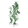
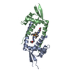
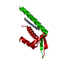
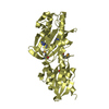
- PDB-2i7s: Crystal structure of Re(phen)(CO)3 (Thr124His)(His83Gln) Azurin C... -
+ Open data

- Basic information
| Entry | Database: PDB / ID: 2i7s | ||||||
|---|---|---|---|---|---|---|---|
| Title | Crystal structure of Re(phen)(CO)3 (Thr124His)(His83Gln) Azurin Cu(II) from Pseudomonas aeruginosa | ||||||
 Components Components | Azurin | ||||||
 Keywords Keywords | ELECTRON TRANSPORT / Azurin / Rhenium / Electron Transfer in proteins | ||||||
| Function / homology |  Function and homology information Function and homology informationtransition metal ion binding / periplasmic space / electron transfer activity / copper ion binding / zinc ion binding / identical protein binding / plasma membrane Similarity search - Function | ||||||
| Biological species |   Pseudomonas aeruginosa (bacteria) Pseudomonas aeruginosa (bacteria) | ||||||
| Method |  X-RAY DIFFRACTION / SYNCHROTRON / MOLECULAR REPLACEMENT / Resolution: 1.35 Å X-RAY DIFFRACTION / SYNCHROTRON / MOLECULAR REPLACEMENT / Resolution: 1.35 Å | ||||||
 Authors Authors | Gradinaru, C. / Crane, B.R. | ||||||
 Citation Citation | Journal: J.Am.Chem.Soc. / Year: 2009 Title: Relaxation dynamics of Pseudomonas aeruginosa Re(I)(CO)3(alpha-diimine)(HisX)+ (X = 83, 107, 109, 124, 126)Cu(II) azurins. Authors: Blanco-Rodriguez, A.M. / Busby, M. / Ronayne, K. / Towrie, M. / Gradinaru, C. / Sudhamsu, J. / Sykora, J. / Hof, M. / Zalis, S. / Di Bilio, A.J. / Crane, B.R. / Gray, H.B. / Vlcek, A. | ||||||
| History |
|
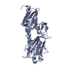
- Structure visualization
| Structure viewer | Molecule: MolmilJmol/JSmol |
|---|
- Downloads & links
-Download
| PDBx/mmCIF format | 2i7s.cif.gz | 185.4 KB | Display | PDBx/mmCIF format |
|---|---|---|---|---|
| PDB format | pdb2i7s.ent.gz | 149.3 KB | Display | PDB format |
| PDBx/mmJSON format | 2i7s.json.gz | Tree view | PDBx/mmJSON format | |
| Others |  Other downloads Other downloads |
-Validation report
| Arichive directory | https://data.pdbj.org/pub/pdb/validation_reports/i7/2i7sftp://data.pdbj.org/pub/pdb/validation_reports/i7/2i7s | HTTPS FTP |
|---|
-Related structure data
| Related structure data |  3iboC  1bexS S: Starting model for refinement C: citing same article ( |
|---|---|
| Similar structure data |


-Links
 PDBj
PDBj- Assembly
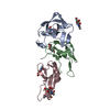
| Deposited unit | 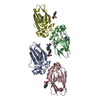
| ||||||||
|---|---|---|---|---|---|---|---|---|---|
| 1 | 
| ||||||||
| 2 | 
| ||||||||
| Unit cell |
|
-Components
| #1: Protein | Mass: 13987.839 Da / Num. of mol.: 4 / Mutation: H83Q, T124H Source method: isolated from a genetically manipulated source Details: Rhenium-modified azurin / Source: (gene. exp.) Pseudomonas aeruginosa (bacteria) / Gene: azu / Species (production host): Escherichia coli / Production host: #2: Chemical | ChemComp-CU /   Mass: 63.546 Da / Num. of mol.: 4 / Source method: obtained synthetically / Formula: Cu Mass: 63.546 Da / Num. of mol.: 4 / Source method: obtained synthetically / Formula: Cu#3: Chemical | ChemComp-REP / ( 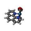  Mass: 450.443 Da / Num. of mol.: 4 / Source method: obtained synthetically / Formula: C15H8N2O3Re Mass: 450.443 Da / Num. of mol.: 4 / Source method: obtained synthetically / Formula: C15H8N2O3Re#4: Chemical | ChemComp-CON / |   Mass: 127.055 Da / Num. of mol.: 1 / Source method: obtained synthetically / Formula: CoH12N4 Mass: 127.055 Da / Num. of mol.: 1 / Source method: obtained synthetically / Formula: CoH12N4#5: Water | ChemComp-HOH / |  Mass: 18.015 Da / Num. of mol.: 464 / Source method: isolated from a natural source / Formula: H2O Mass: 18.015 Da / Num. of mol.: 464 / Source method: isolated from a natural source / Formula: H2OHas protein modification | Y | |
|---|
-Experimental details
-Experiment
| Experiment | Method: X-RAY DIFFRACTION / Number of used crystals: 1 |
|---|
- Sample preparation
| Crystal | Density Matthews: 2.19 Å3/Da / Density % sol: 43.86 % |
|---|---|
| Crystal grow | Temperature: 298 K / Method: vapor diffusion, hanging drop / pH: 7 Details: 20% PEG4000, 100 mM LiNO3, 100 mM Imidazole pH 7.0. One fourth of the drop volume was saturated with [Co(NH3)5Cl]Cl2 solution, VAPOR DIFFUSION, HANGING DROP, temperature 298.0K |
-Data collection
| Diffraction | Mean temperature: 100 K |
|---|---|
| Diffraction source | Source: SYNCHROTRON / Site: CHESS  / Beamline: F2 / Wavelength: 0.916 Å / Beamline: F2 / Wavelength: 0.916 Å |
| Detector | Type: ADSC QUANTUM 210 / Detector: CCD / Date: Feb 1, 2003 |
| Radiation | Monochromator: Si 111 CHANNEL / Protocol: SINGLE WAVELENGTH / Monochromatic (M) / Laue (L): M / Scattering type: x-ray |
| Radiation wavelength | Wavelength: 0.916 Å / Relative weight: 1 |
| Reflection | Resolution: 1.35→30 Å / Num. all: 171226 / Num. obs: 104393 / % possible obs: 95.6 % / Observed criterion σ(I): 2 / Redundancy: 1.64 % / Rsym value: 0.08 / Net I/σ(I): 4.7 |
- Processing
| Software |
| ||||||||||||||||||||||||||||||||
|---|---|---|---|---|---|---|---|---|---|---|---|---|---|---|---|---|---|---|---|---|---|---|---|---|---|---|---|---|---|---|---|---|---|
| Refinement | Method to determine structure: MOLECULAR REPLACEMENT Starting model: PDB entry 1BEX Resolution: 1.35→30 Å / Isotropic thermal model: anisotropic / Cross valid method: FREE-R / σ(I): 2 / Stereochemistry target values: Engh & Huber Details: LEAST-SQUARES ANISOTROPIC REFINEMENT USING THE KONNERT-HENDRICKSON CONJUGATE-GRADIENT ALGORITHM
| ||||||||||||||||||||||||||||||||
| Displacement parameters | Biso mean: 26.211 Å2 | ||||||||||||||||||||||||||||||||
| Refinement step | Cycle: LAST / Resolution: 1.35→30 Å
|
